39 correctly label the following anatomical features of the stomach wall.
› manual › 1548521PHILIPS EPIQ 7 USER MANUAL Pdf Download | ManualsLib In addition, you should do the following: • Consider the patient’s size and ability to accommodate the transducer tip and shaft. EPIQ 7 User Manual 4535 617 25341... Page 224: Patient Selection For Tee Transducer Use The following table lists the minimum patient weight recommendations when using TEE transducers. Antenatal Care Module: 3. Anatomy and Physiology of the Female ... Behind (posterior to) the uterus, cervix and vagina in Figure 3.1, you can see part of the large intestine (colon) and the rectum, where solid waste is directed out of the body through the anus. Knowing about the anatomical position of all these structures is very important during pregnancy, labour and delivery.
The Anatomical Regions of the Body - dummies The anatomical regions (shown) compartmentalize the human body. Just like on a map, a region refers to a certain area. The body is divided into two major portions: axial and appendicular. The axial body runs right down the center (axis) and consists of everything except the limbs, meaning the head, neck, thorax (chest and back), abdomen, and pelvis.
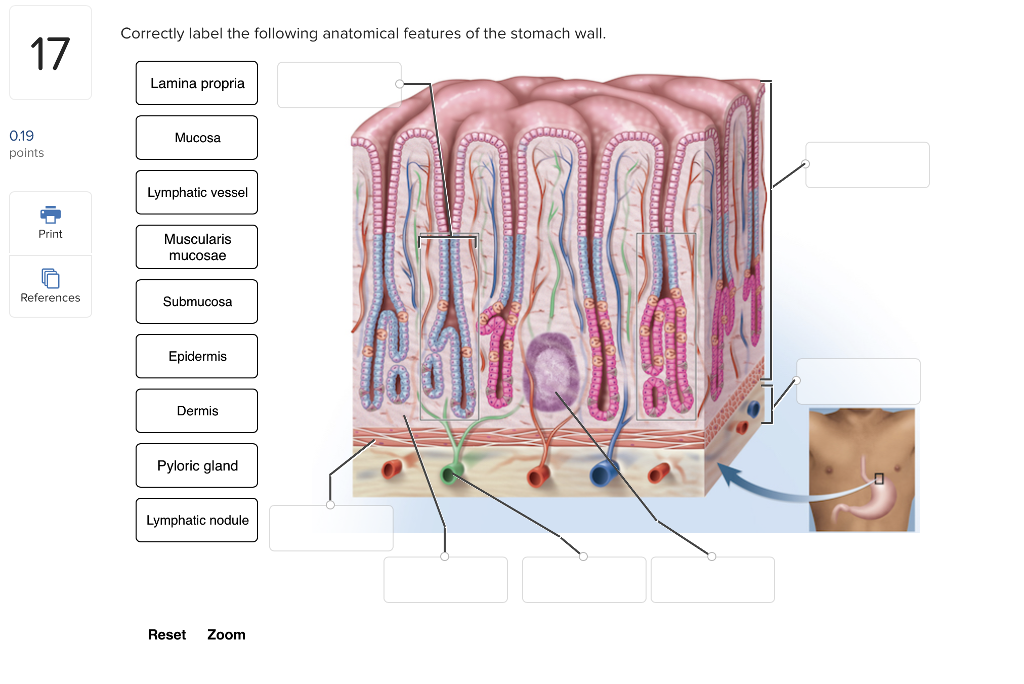
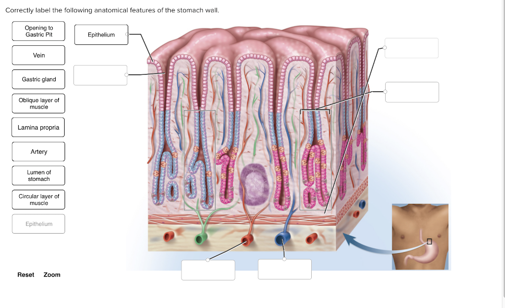
Correctly label the following anatomical features of the stomach wall.
thoracic cavity | Description, Anatomy, & Physiology | Britannica thoracic cavity, also called chest cavity, the second largest hollow space of the body. It is enclosed by the ribs, the vertebral column, and the sternum, or breastbone, and is separated from the abdominal cavity (the body's largest hollow space) by a muscular and membranous partition, the diaphragm. It contains the lungs, the middle and lower airways—the tracheobronchial tree—the heart ... (Get Answer) - Digestive Lab Worksheet i Saved Correctly label the ... Digestive Lab Worksheet i Saved Correctly label the cells found in the stomach 18 Mucous neck cell 0.19 points Hepatocyte Lymphocyte ERRRRRRRRR eBook Mucous cell Print Chief cell References Regenerative cell G cel Parietal cell Reset Zoom Digestive Lab Worksheet 6 Correctly label the following anatomical features of the stomach wall 19 Mucous cell 0.19 Gastric gland CORO points Lymphatic ... Fountain Essays - Your grades could look better! We offer assignment help in more than 80 courses. We are also able to handle any complex paper in any course as we have employed professional writers who are specialized in different fields of study. From their experience, they are able to work on the most difficult assignments. The following are some of the course we offer assignment help in ...
Correctly label the following anatomical features of the stomach wall.. Correctly label the following anatomical features of the stomach wall ... Diagram | Quizlet Correctly label the following anatomical features of the stomach wall. STUDY Learn Write Test PLAY Match + − Created by Sarah_Branning Terms in this set (6) Epithelium ... Opening to gastric pit ... Lamina propria ... Lamina propria ... vein ... artery ... Practice Quiz - Abdominal Wall - Anatomy Site The normal pattern of venous and lymphatic drainage of the superficial tissues of the anterior abdominal wall is arranged around a horizontal plane. Above that plane, drainage is in a cranial direction; below the plane drainage is in a caudal direction. This reference plane corresponds to: Transpyloric plane Level of anterior superior iliac spines The microscopic anatomy of the esophagus including the individual ... The esophagus also has unique features such as patches of gastric mucosa called inlet patches at the very proximal part and it has a special sphincter mechanism at the most distal aspect. This review covers the normal microscopic anatomy of the esophagus and the patterns of reaction to stress and injury of each layer and each special structure. Solved Correctly label the following anatomical features of - Chegg Question: Correctly label the following anatomical features of the stomach wall. Opening to Gastric Pit Epithelium Vein Gastric gland HINCHABERLU LLLLAA LLLLL IRRARA HANSEN ?????????????????
Chapter 8. Serous Membranes of the Abdominal Cavity The neurovascular and lymphatic supply of the peritoneum course to and from the posterior abdominal wall and gut tube through the two-layered mesentery (Figure 8-1B).The vascular supply to the parietal peritoneum is through the same vessels that supply the abdominal body wall, mainly the intercostal, lumbar, and epigastric vessels.The vascular supply to the visceral peritoneum is through ... Abdominal wall: Layers, muscles and fascia | Kenhub The abdominal wall surrounds the abdominal cavity, providing it with flexible coverage and protecting the internal organs from damage. It is bounded superiorly by the xiphoid process and costal margins, posteriorly by the vertebral column and inferiorly by the pelvic bones and inguinal ligament . Thorax: Anatomy, wall, cavity, organs & neurovasculature | Kenhub The chest, properly called the thorax, is the superior part of the trunk located between the neck and abdomen. It consists of several components: Thoracic wall Several cavities Neurovasculature and lymphatics Internal organs Breasts On this page, we'll briefly take a look at each of the above components and how they fit together to form the thorax. Chapter 25 HW Flashcards - Quizlet Correctly label the following anatomical features of the stomach wall (2) Correctly label the cells found in the stomach. Correctly label the following microscopic anatomy of the liver. Correctly label the following parts of intestinal villi. Click and drag the labels to match each enzyme with its function.
ReaderUi ReaderUi villus | anatomy | Britannica The inner wall of the small intestine is covered by numerous folds of mucous membrane called plicae circulares. The surface of these folds contains tiny projections called villi and microvilli, which further increase the total area for absorption. Absorbed nutrients are moved into circulation by blood capillaries and lacteals, or lymph channels. The Peritoneum - Visceral - Parietal - TeachMeAnatomy The abdominal viscera can be divided anatomically by their relationship to the peritoneum. There are two main groups: intraperitoneal and retroperitoneal organs. Intraperitoneal Organs Intraperitoneal organs are enveloped by visceral peritoneum, which covers the organ both anteriorly and posteriorly. Examples include the stomach , liver and spleen. Chapter 24 Digestive System Flashcards - Quizlet Correctly label the anatomical features of a ... Test your knowledge about the esophagus by correctly indicating which of the following are ... intestine include those that produce mucus which aids in protecting it from the digestive enzymes and acid from the stomach. Bound to the inner wall of the intestines are disaccharidases and peptidases ...
Solved: Correctly Label The Following Anatomical Features ... | Chegg.com
quizlet.com › 445722440 › anatomy-midterm-lectureAnatomy Midterm Lecture Flashcards - Quizlet Correctly label the following anatomical features of the heart and thoracic cage. Correctly label the following structures related to the position of the heart in the thorax. Correctly label the following parts of the pericardium and the heart walls
Solved: Correctly Label The Following Anatomical Features ... | Chegg.com
23.4 The Stomach - Anatomy & Physiology First, the stomach wall is covered by a thick coating of bicarbonate-rich mucus. This mucus forms a physical barrier, and its bicarbonate ions neutralize acid. Second, the epithelial cells of the stomach's mucosa meet at tight junctions, which block gastric juice from penetrating the underlying tissue layers.
Post a Comment for "39 correctly label the following anatomical features of the stomach wall."