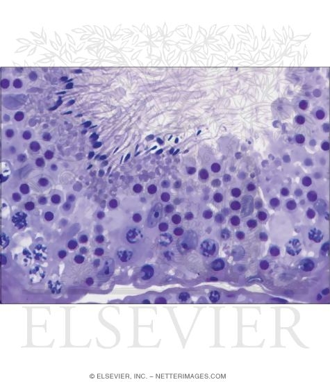
40 label the light micrograph of the seminiferous tubule using the hints provided
[Solved] Label the micrograph of the renal corpuscle and surrounding ... Answer to Label the micrograph of the renal corpuscle and surrounding structures using the hints. Study Resources. Main Menu; ... Label the micrograph of the renal corpuscle and surrounding structures using the hints provided. Proximal convoluted tubule Capsular space Macula densa Bowman's capsule (parietal laver) Distal convoluted tubule ... Seminiferous Tubules, Light Micrograph IPhone 12 Case Seminiferous Tubules, Light Micrograph IPhone 12 Case for Sale by Steve Gschmeissner. Protect your iPhone 12 / 12 Pro with an impact-resistant, slim-profile, hard-shell case. The image is printed directly onto the case and wrapped around the edges for a beautiful presentation. Simply snap the case onto your iPhone 12 / 12 Pro for instant protection and direct access to all of the phone's features!
Seminiferous tubules, light micrograph - Stock Image - F005/7287 Seminiferous tubules. Coloured light micrograph of a section through the testis, showing seminiferous tubules (pink) and lending cells (yellow). This is the site of sperm production (spermatogenesis). Magnification: x150 when printed at 10 centimetres wide.

Label the light micrograph of the seminiferous tubule using the hints provided
Seminiferous Tubules, Light Micrograph by Steve Gschmeissner Seminiferous Tubules, Light Micrograph is a photograph by Steve Gschmeissner which was uploaded on May 6th, 2013. The photograph may be purchased as wall art, home decor, apparel, phone cases, greeting cards, and more. All products are produced on-demand and shipped worldwide within 2 - 3 business days. Seminiferous tubules. Light micrograph of a cross- section (Photos ... Seminiferous tubules. Light micrograph of a cross- section through seminiferous tubules in an earthworm (Lumbricus terrestris) testis. The seminiferous tubules are the site of sperm production (spermatogenesis). In the central region of each tubule are spermatozoa, formed from gametes undergoing initial meiosis and later spermiogenesis. Ch 27 Lab Flashcards - Quizlet Label the testis and spermatic cord using the hints provided. 1. The ovaries are paired, oval-shaped organs found in the pelvic cavity. 2. The uterine tubes receive the ovulated oocyte and are the most common place for fertilization to take place. 3. The uterus is a pear shaped muscular organ where fetal development takes place. 4.
Label the light micrograph of the seminiferous tubule using the hints provided. Seminiferous Tubule, Light Micrograph by Steve Gschmeissner Seminiferous Tubule, Light Micrograph is a photograph by Steve Gschmeissner which was uploaded on May 6th, 2013. The photograph may be purchased as wall art, home decor, apparel, phone cases, greeting cards, and more. All products are produced on-demand and shipped worldwide within 2 - 3 business days. Label the light micrograph of the seminiferous tubule using the hints ... Label the light micrograph of the seminiferous tubule using the hints provided STUDY Learn Write Test PLAY Match Created by Sarah_Branning Terms in this set (4) lumen ... spermatid ... germ cell ... seminferous tubuke ... Focus on some of the seminiferous tubules using high Focus on some of the seminiferous tubules using high. School Kaplan University, Cedar Rapids; Course Title MEDICAL AS 121; Type. Homework Help. Uploaded By akbarnett. Pages 10 Ratings 94% (18) 17 out of 18 people found this document helpful; This preview shows page 4 - 9 out of 10 pages. ... Light Micrograph of the Wall of a Seminiferous Tubule Please Note: You may not embed one of our images on your web page without a link back to our site. If you would like a large, unwatermarked image for your web page or blog, please purchase the appropriate license.
Seminiferous tubule, light micrograph by Science Photo Library Seminiferous tubule, light micrograph is a photograph by Science Photo Library which was uploaded on March 7th, 2014. The photograph may be purchased as wall art, home decor, apparel, phone cases, greeting cards, and more. All products are produced on-demand and shipped worldwide within 2 - 3 business days. Solved Label the micrograph of the urinary bladder using the | Chegg.com Science; Anatomy and Physiology; Anatomy and Physiology questions and answers; Label the micrograph of the urinary bladder using the hints provided 26 8 00:33 Submucosa Lamina propria Transitional epithelium Detrusorm CMTV Waco Teiser Reset Zoom Which structure is highlighted? 25 8003121 Multiple Choice external urethral sphincter Internal urethral sphincter external urethral orice ureter O ... Make a drawing of the seminiferous tubule and label the structures Let me describe the structure of seminaries to view seminar first rib you'll are lined by two types off settles that is spermatozoa Gornje and certain light cells from inside spermatozoa Gornje cells farms bombs through my topic. Division through my daughter. Take division. Why nutrition to germ cells, nutrition too? Job cells is provided by certain license. Light Micrograph of a Seminiferous Tubule In Transverse Section Light Micrograph of a Seminiferous Tubule In Transverse Section. Variant Image ID: 14649. Add to Lightbox. Save to Lightbox. Email this page. Link this page. Print. Please describe! how you will use this image and then you will be able to add this image to your shopping basket.
Seminiferous tubules, light micrograph Stock Photo | u18309637 | Fotosearch Seminiferous tubules, light micrograph Stock Photo - Lushpix. u18309637 Fotosearch Stock Photography and Stock Footage helps you find the perfect photo or footage, fast! We feature 64,900,000 royalty free photos, 337,000 stock footage clips, digital videos, vector clip art images, clipart pictures, background graphics, medical illustrations, and maps. Seminiferous tubule, light micrograph - Yard Sign | CafePress Shop Seminiferous tubule, light micrograph - Yard Sign designed by Science-Photo-Library. Lots of different size and color combinations to choose from. Free Returns High Quality Printing Fast Shipping Seminiferous tubule, light micrograph Poster by Science Photo Library Seminiferous tubule, light micrograph Poster by Science Photo Library. All posters are professionally printed, packaged, and shipped within 3 - 4 business days. Choose from multiple sizes and hundreds of frame and mat options. 25% off all wall art! It's our once-a-year wall art sale! Ch 27 Reproductive Flashcards | Quizlet Cells arrested in metaphase, when chromosomes are most highly condensed, are stained and then viewed with a microscope equipped with a camera. A photo is displayed and the images of the chromosomes are arranged into pairs according to their appearance. • This karyotype shows the chromosomes from a normal human male. Homologous Chromosomes/Homologs
Light microscope view of a seminiferous tubule in the Pelamis platarus ... Download scientific diagram | Light microscope view of a seminiferous tubule in the Pelamis platarus testis. The seminiferous tubule has a wide lumen and thick germinal epithelium containing ...
Post a Comment for "40 label the light micrograph of the seminiferous tubule using the hints provided"