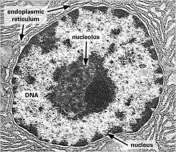
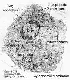
40 label the transmission electron micrograph of the cell
Transmission Electron Microscopy Cell [Transmission Electron Microscopy Cell] - 8 images - electron microscopy auditory science lab, Three-dimensional analysis of interstitial cells in the lamina propria ... The connective connection: Interstitial cells of Cajal (ICC) and ICC-like cells establish synapses with immunoreactive cells: Electron microscope study in situ. J. Cell. Mol. Med. 9, 714-730 (2005).
Nanomaterials | Free Full-Text | Dendritic Mesoporous Silica Hollow ... The high-angle annular dark field scanning transmission electron microscopy (HAADF-STEM) image and energy dispersive X-ray spectroscopy (EDX) element mappings were achieved by the FEI instrument. X-ray diffraction (XRD) patterns were obtained using Cu-K radiation (λ = 1.54056 Å) with a tube voltage of 5 kV and a current of 30 mA on a Bruker ...

Label the transmission electron micrograph of the cell
Modified dendritic cell-derived exosomes activate both NK cells and T ... NK cells or T lymphocytes were coincubated with CML-RAE-1γ-Dex vaccines. Flow cytometry was performed to evaluate the activation and proliferation of these immune cells. ... -coated copper grids and then negatively stained with 2% uranyl acetate. Ultimately, exosomal morphology was imaged by transmission electron microscopy (TEM, Tecnai G2 ... Mitochondrion - Wikipedia The inner mitochondrial membrane contains proteins with three types of functions: Those that perform the electron transport chain redox reactions ATP synthase, which generates ATP in the matrix Specific transport proteins that regulate metabolite passage into and out of the mitochondrial matrix Immunofluorescence-Mediated Detection of Respiratory Virus Infections ... Tapping the flask against the palm of your hand may help to release the cells. 16. Add 10 ml of the trypsin inhibitor solution (TIS). 17. Gently homogenize the cell suspension using a serological pipette, then transfer the cell suspension to a 50-ml conical tube. 18. Centrifuge for 5 min at 350 × g, room temperature. 19.
Label the transmission electron micrograph of the cell. High-Pressure Freezing and Transmission Electron Microscopy to ... Transmission electron microscopy (TEM) is the main technique used to study the ultrastructure of biological samples. Chemical fixation was considered the main method for preserving samples for TEM; however, it is a relatively slow method of fixation and can result in morphological alterations. Global Microscopy Device Market - 2022-2029 Electron microscopy is a technique for obtaining high-resolution images at the speed of biological as well as non-biological samples. It is one of the most important techniques available for the analysis of properties such as morphology, crystal and defect structure, elemental composition, and electronics of any kind sample. Unraveling the mechanism for paired electrocatalysis of organics with ... Scanning electron microscopy (SEM) and transmission electron microscopy (TEM) images of FeP-MoP/FF reveal a coarse nanosheet with the size and thickness of 500 and 50 nm, respectively (Fig. 1b, c... Transmission Electron Microscopy Cell [Transmission Electron Microscopy Cell] - 8 images - in almost living color the first colored electron micrographs of cells,
Plasmodium infection suppresses colon cancer growth by inhibiting ... Transmission electron microscopy (TEM) was used to observe the ultrastructural change in colon cancer cells, and the expression of mitochondrial biogenesis correlative central protein, PGC-1α, and mitophagy relevant crucial proteins, PINK1/Parkin, were detected by western blot. Results Minimalist O2 generator formed by in situ KMnO4 oxidation for tumor ... We then characterized the morphology and size of our obtained MnO 2 products with transmission electron microscopy (TEM), scanning electron microscopy (SEM), and dynamic light scattering (DLS). The prepared BSA-MnO 2 and DNA-MnO 2 have a sphere-like geometry and good monodispersity ( Figure S9a,b ), and the hydrodynamic sizes achieved in ... Alternative splicing encodes functional intracellular CD59 isoforms ... transmission electron microscopy with immunogold labeling for iris-1 or iris-2 (20 nm gold particles), and insulin (10 nm gold particles), showed that the level of colocalization between iris isoforms and insulin granules increased in high glucose conditions, consistent with cd59-iris isoforms being involved in insulin secretion ( fig. 2 a and b, … Plant biology: Peering deeply into the structure of the ... - cell.com Cell wall ultrastructure has previously been assessed by thin-section transmission electron microscopy and by surface-based methods, such as atomic force microscopy. A new study uses electron tomography to image cellulose and pectin organization deep inside a thick epidermal cell wall.
Distribution of interstitial cells of Cajal in the Esophagus and change ... Transmission electron microscope (TEM) Tissues were cut from the upper, middle, and lower parts of the esophagus, fixed, and embedded in 2.5% glutaraldehyde solution overnight. Subsequently, the specimens were immersed in 1% osmium acid solution at 4 °C for 2 h, following which they were immersed in 2% uranyl acetate solution at 37 °C for 1 h. 2 monkeypox strains in US suggest possible undetected spread WHO Monkeypox FILE - This 2003 electron microscope image made available by the Centers for Disease Control and Prevention shows mature, oval-shaped monkeypox virions, left, and spherical immature ... Engineered Exosomes Loaded with miR-563 Inhibit Lung Cancer Growth Exosomes were viewed with a transmission electron microscope using Jeol JEM-1400 (Jeol Ltd., Tokyo, Japan) operating at 80 kV. The size of exosomes was analyzed using a Zetasizer Nano ZS system (Malvern Instruments, Malvern, UK). 2.12. Western Blot Mechanistic Approach of Nano Carriers for Targeted in Cancer ... Transmission Electron Microscopy (TEM) The morphology of nanolipid vesicles was detected using a transmission electron microscope [FEI (Type FP 5018/40) Tecnai G2 Spirit Bio TWIN]. The formulations were dispersed in de-ionized water at a concentration of 500 μg/mL.
FluoroNanogold: Fluorescence and Electron Micrographs of Labeled SC35 ...
Boosting Electrocatalytic CO2 Reduction with Conjugated Bimetallic Co ... Transmission electron microscopy (TEM) images were recorded by a FEI Tecnai 20 microscope (FEI, United States) at 200 kV. The energy-dispersive system (EDS) of samples were performed with a Titan Cubed Themis G2 300 high-resolution transmission electron microscope (FEI, United States) operated at 200 kV.
Cells Formative Assignment
Histological Injury to Rat Brain, Liver, and Kidneys by Gold ... Transmission electron microscopy (TEM) was used to determine the size, size distribution, and shape of GNPs. ... Colloidal gold nanoparticles are found in dispersed and aggregated forms within the cell cytoplasm and provide anat. labeling information, but their uptake is nonspecific for malignant cells. The anti-EGFR antibody conjugated ...

Smell Receptors Transmission Electron Micrograph Stock Illustration ...
Alternative splicing of BUD13 determines the severity of a ... Transmission electron microscopy Cells were grown on Thermanox plastic coverslips (Thermo Fisher Scientific) and fixed in 2.5% glutaraldehyde. Cells were stained in 0.5% (volume/volume) osmium tetroxide (Science Services) and contrasted in 0.1% (weight/volume) tannin and 2% (weight/volume) uranyl acetate (Sigma-Aldrich, Merck).
Basic anatomy
Which infectious agent causes tuberculosis (TB) (consumption)? Under a high magnification of 15549x, this scanning electron micrograph depicts some of the ultrastructural details seen in the cell wall configuration of a number of Gram-positive Mycobacterium...
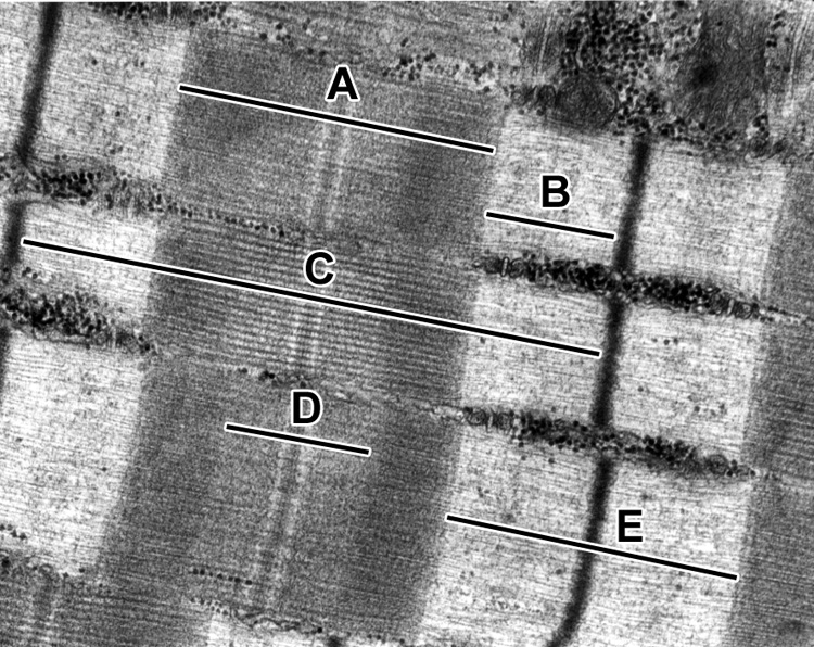
Muscle | histology
How Food Microscopy Determines Nutritional Quality of Cereals Fluorescence microscopy is a commonly chosen technique to look for the presence of adulterants or contaminants in a food sample. Physical contaminants such as glass, stones or dirt can be...
Topic 1.2 Ultra-Structure of Cells - AMAZING WORLD OF SCIENCE WITH MR ...
Antibiotic - Wikipedia An antibiotic is a type of antimicrobial substance active against bacteria.It is the most important type of antibacterial agent for fighting bacterial infections, and antibiotic medications are widely used in the treatment and prevention of such infections. They may either kill or inhibit the growth of bacteria. A limited number of antibiotics also possess antiprotozoal activity.
Hermaphrodite Coelomocyte System
51 Postdoctoral Fellowship at National University of Singapore (NUS ... The postdoctoral fellow will study ovarian ageing via 1) tissue characterization (e.g., electron microscopy, mass spectrometry), 2) development of instructive biomaterials for in vitro cell culture, and 3) elucidation of mechanosensitive signaling pathways contributing to functional decline. Unique proposals related to the study of ovarian ...
The Cell: The Histology Guide
What is the pathophysiology of hepatitis B (HBV) (Hep B ... - Medscape The viral genome is a partially double-stranded, circular DNA linked to a DNA polymerase that is surrounded by an icosahedral nucleocapsid and then by a lipid envelope. Embedded within these layers...
monograph 1
Combination of Live Cell Surface-Enhanced Raman Scattering Imaging with ... SERS tags can be used as nontoxic cell labeling moieties or as indirect sensors to detect the presence of biomolecules. One of the benefits of SERS microscopy over, for ... TEM images were collected with a JEOL JEM-1400PLUS transmission electron microscope operating at 120 kV, using carbon-coated 400 square mesh copper grids. ...
Post a Comment for "40 label the transmission electron micrograph of the cell"