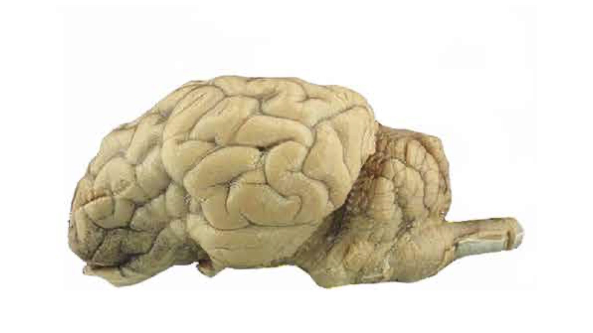
38 sheep brain unlabeled
heart diagram unlabeled heart anatomy sheep dissection circulatory artery biologycorner brachiocephalic valve semilunar system diet aortic apex recipes 4d tendineae chordae. Posterior View Of Surface Anatomy Of Heart Quiz . posterior heart anatomy surface quiz human gross brain purposegames. Human Heart Diagram - Unlabeled - Tim's Printables Label the parts of a Sheep Brain (Midsagittal) Quiz Label the parts of a Sheep Brain (Midsagittal) a quiz by Dr. Smith's BSC 2085L. •.
diagram of brain labeled brain diagram unlabeled human half anatomy clipart left blank clip cliparts labeled psych ap results etc clipartmag tiff usf edu. 25. Midsagittal Sheep Brain (labeled) | Anatomyphyslab261 | Flickr . midsagittal. TooSogiE Medical Images: Cranial Nerves : X - XII toosogie-medical-images.blogspot.com.

Sheep brain unlabeled
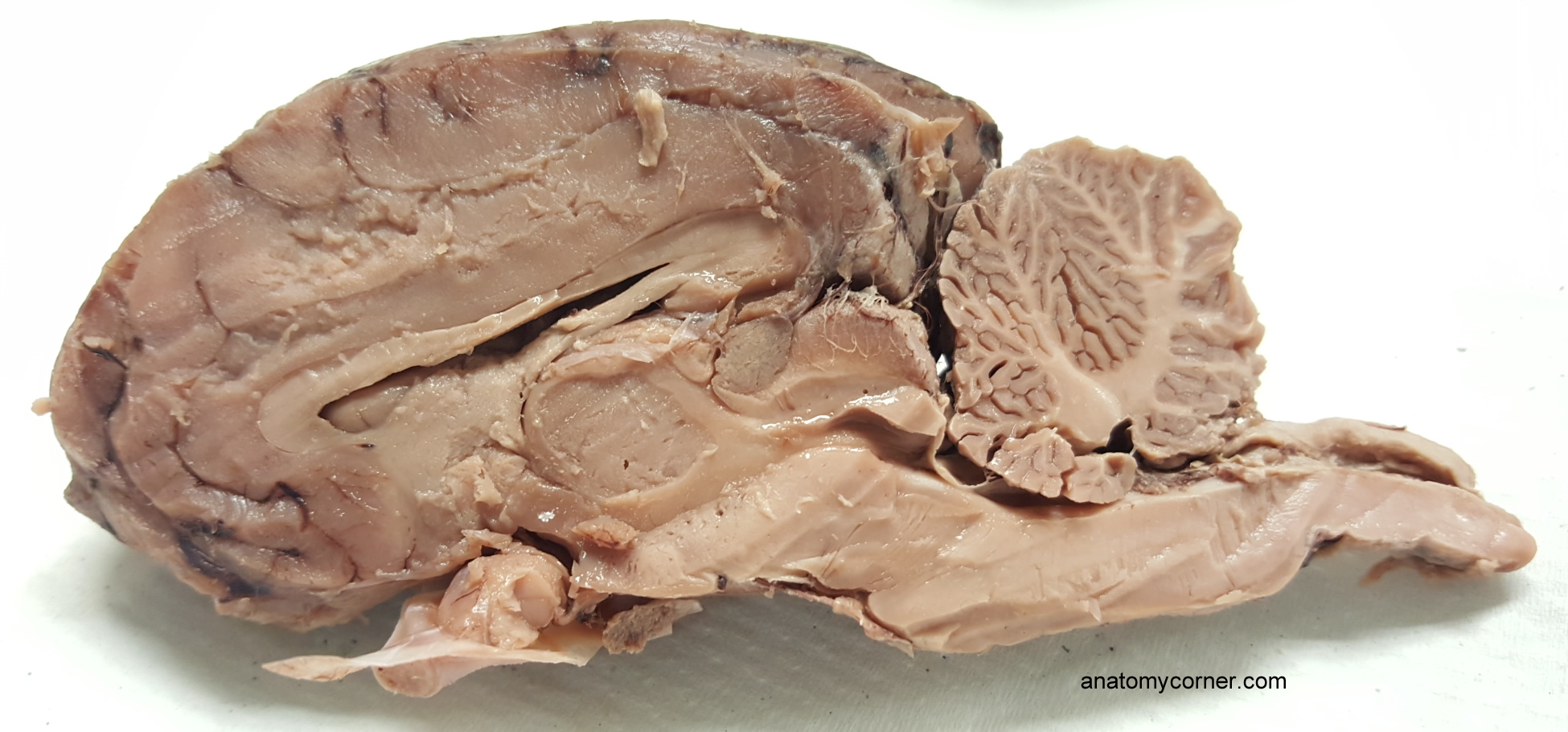
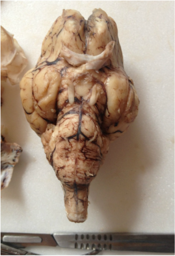
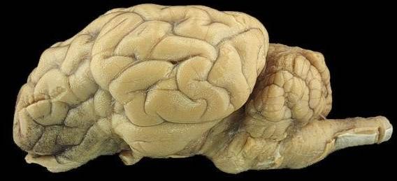
Sheep Brain - midsagittal Flashcards | Quizlet Exam 1: Brain Models and Sheep Brain III. 33 terms. kyle_thun. Cerebrum. 22 terms. Jessica_Turner51 PLUS. Respiratory. 47 terms. dezirae-brian_hays. Anatomy Lab: Nervous System. 86 terms. sreisaacs. Other sets by this creator. Intro to Oncology - Drugs. 21 terms. ryankidd01. STLCOP - APPE 2016 - Unit #9 Brand Name. 20 terms. Sheep Brain - Dorsal View The rostral colliculus(large arrow label) and the caudal colliculus(small arrow label) together form the tectumof the midbrain. Also labeled are the pineal body(green), the caudate nucleus(1), the floor of the fourth ventricle(white and pink) and cerebellar peduncles(blue = rostral, red = middle, and yellow = caudal). Go Top Sheep Brain Dissection Project Guide | HST Learning Center Place the brain with the curved top side of the cerebrum facing up. Use a scalpel (or sharp, thin knife) to slice through the brain along the center line, starting at the cerebrum and going down through the cerebellum, spinal cord, medulla, and pons. Separate the two halves of the brain and lay them with the inside facing up. 2.
Sheep brain unlabeled. the brain diagram labeled sheep brain dissection parker dr Pin On Unlabeled Anatomy nervous system unlabeled Human Circulatory System Diagram Labeled Circulatory System The Free heart diagram human circulatory labeled system simple anatomy circulation attack notes Click On The Links Below To See Labeled Images Of A Dissected Pigeon Sheep brain dissection | Human Anatomy and Physiology Lab (BSB 141 ... Examining the external sheep brain. The tough outer covering of the sheep brain is the dura mater, the outermost meninges membrane covering the brain.Remove the dura mater to see most of the structures of the brain, but remove it carefully, so as to leave all the other structures beneath it intact. Removing the dura mater from the cerebellum at the back of the brain can be tricky. Sheep Brain Instructions - University of Scranton random plate selection (also, to test yourself) This button allows you to toggle between labeled and unlabeled images. On many of the images you will see brackets such as the ones below. A bracket of this type is used to designate an area or region of the brain. Brackets, or lines, which end in small circles designate hollow structures. Sheep Brain Labeling (part 1) Quiz - By dilatory - Sporcle Sheep Brain Labeling (part 1) Quiz - By dilatory. Science sheep. QUIZ LAB SUBMISSION. Random Science or sheep Quiz.
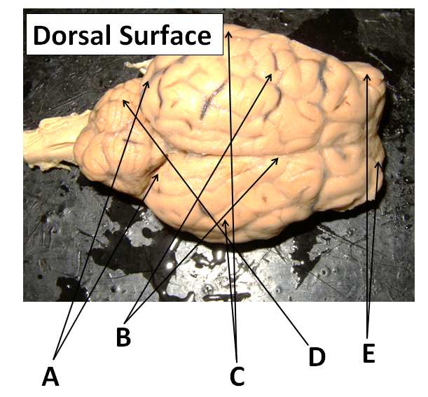
anatomy of a sheep Vintage Sheep Clip Art - White And Black - The Graphics Fairy thegraphicsfairy.com. sheep clip graphics fairy. BIO201-Sheep Brain savalli.us. brain sheep frontal section labeled bio201 savalli unlabeled return. WMU Psychology Department: Lisa Baker homepages.wmich.edu. brain sheep nerves cranial ventral psychology wmich homepages edu. Sheep kidney. Sheep Brain Dissection with Labeled Images - The Biology Corner 1. The sheep brain is enclosed in a tough outer covering called the dura mater. You can still see some structures on the brain before you remove the dura mater. Take special note of the pituitary gland and the optic chiasma. These two structures will likely be pulled off when you remove the dura mater. Brain with Dura Mater Intact Sheep Brain Neuroanatomy Online Self-Test | KPU.ca - Kwantlen ... Sheep Brain Neuroanatomy Online Self-Test Use each diagram as a reference, and selected the correct answer for each lettered structure. You may find it useful to open the diagrams in a separate window to review while answering each question. Dorsal Surface Dorsal Surface A * Occipital Lobe Temporal Lobe Cerebellum Parietal Lobe Dorsal Surface B * Practice Lab Practical on the Sheep Brain Identify the cleft labeled 7. Look here for the answer Transverse fissure Identify the shiny membrane visible on the sheep brain surface. Look here for the answer Pia mater In the above picture: Identify the structure labeled 1. Look here for the answer Olfactory bulb Identify the structure labeled 2. Look here for the answer
Brain Labeling Worksheet brain anatomy sheep dissection label unlabeled worksheet lab sheet systems body biology labeled guide ap biologycorner parts lateral psychology practice. Sheep Brain Dissection @ Fort Vancouver! - NW NOGGIN: Neuroscience nwnoggin.org. brain sheep dissection superior vancouver fort skyview storm colliculi prepared handout angela johnson nwnoggin Sheep Heart Images - San Diego Mesa College Sheep Brain Images; Practice Exams; Fetal Pig Images. Fetal Pig Images; Practice Exams; Heart. Heart Models; Sheep Heart Images; Practice Exams; Additional Resources; SHEEP HEART IMAGES. Sheep Heart Unlabeled. Sheep Heart Leader-lined. Sheep Heart Labeled. San Diego Mesa College 7250 Mesa College Drive San Diego, CA 92111-4998 Student Support ... Sheep brain Flashcards | Quizlet Sheep brain Identification of structures observed during sheep brain dissection. STUDY PLAY dura mater Identify the covering. cerebrum Identify the major brain region. cerebellum Identify the major brain region. olfactory bulb Identify the tip. optic nerve Identify the nerve by name. optic chiasma Identify the "x". optic chiasma ventral brain anatomy Brain Stem Unlabeled anatomycorner.com. unlabeled anatomycorner. Print Neuro Lab Block I- Brain Stem Gross Anatomy Flashcards | Easy . ... midsagittal cerebral perspective neurosurgicalatlas correlation. Sheep Brain play.psych.mun.ca. sheep sagittal caudate brain. Control Of Craving By The Prefrontal Cortex | PNAS .
What are the differences between a sheep brain and human brain?
diagram of the human brain Unlabeled Digestive System Diagram - Koibana.info In 2020 | Digestive . digestive system diagram human unlabeled body tract labels organs anatomy koibana info. Sheep Brain Flashcards | Quizlet quizlet.com. sheep brain cerebellum quizlet flashcards. Sheep Brain Dissection Guide | Medicine Studies, Medical Laboratory

Sagittal Brain
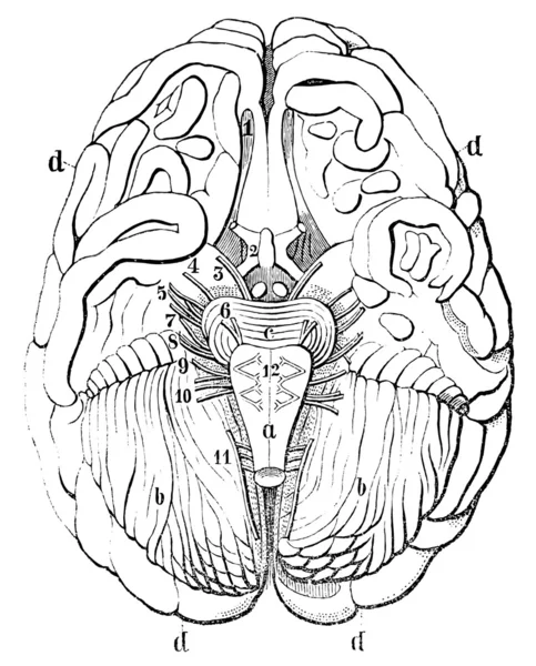
Sheep | isu-bio-351 A = Gyri (ridges) B = Sulcus (fissure) Circled = Cerebral hemisphere. press to zoom. 1/5. Diencephalon ventral view. A = Optic chiasma B = Infundibulum C = Pituitary II = Optic nerve. press to zoom. Diencephalon ventral view. Pituitary removed A = Optic chiasma B = Infundibulum C = Mamillary body Hypothalamus is from the optic chiasm to ...

Sheep Brain Dissection Images
Sheep Brain Label | Dissection, Brain anatomy, Human brain diagram Sheep Brain Label A drawing of the brain with the parts unlabeled. Students can practice naming the parts of the brain, then check their answers with the provided key. Biologycorner 17k followers More information unlabeled brain Find this Pin and more on A&P by Dijana Kovacevic. Brain Gym For Kids Human Brain Diagram Brain Anatomy And Function

Requirements for the Concentration – Neuroscience
PDF Sheep Brain Practical Study Guide - auburn.k12.il.us Sheep Brain Practical Study Guide. Dura Mater. Olfactory Bulb Pituitary Gland Dura Mater Optic Chiasm. Corpus Callosum Longitudinal Fissure Lateral Ventricle Gray Matter White Matter. Arbor Vitae "Tree of Life" Cerebellum "Little Brain" ...

Game Statistics - sheep brain
brain diagram unlabeled Spinal cord anatomy nerves nervous quiz system brain mater autonomic commissure gray nerve exercise quizlet boundary anterior dissection sheep superior Pig Heart Diagram Labeled : Biological Science Picture Directory. 9 Pictures about Pig Heart Diagram Labeled : Biological Science Picture Directory : Pin on unlabeled Anatomy, Midsagittal ...

Eva Pena Burgos di Twitter: "Term newborn 👶🽠who died at 1 ...
internal parts of the brain heart diagram anatomy labeled worksheet physiology animals labelled answers human worksheets system label parts structure simple blood wikieducator body unlabeled. Sheep Brain #2 . sheep brain anatomy label nervous system dissection lateral ventricle section sagittal sdmesa classroom edu cerebral diagram aqueduct lesson ...

Median Sagittal Section of Brain
Lab 9—Sheep Brain—Labeled The Sheep's Brain Return to: The Unlabeled Brains Lab 9 Page BIO 137 Main Page Be sure to practice identifying the structures using the unlabeled photos. This page created and maintained by Udo M. Savalli. Last updated August 13, 2005. ...
SCB209 - Lab2 - Natural Sciences Open Educational Resources
Unlabeled Sheep Brain Dissection Images and Link (1).pptx Labs 5&6 Nervous System Whiting Online.doc. University of Pennsylvania. BIOL 109
Lab - Sheep Brain: MAH-Summer 2019-Anatomy and Physiology I
BIO201-Sheep Brain This page last updated 18 August 2019 by Udo M. Savalli ()Images and text © Udo M. Savalli. All rights reserved.
1 Dissection of the Sheep Brain Applications
human heart diagram unlabeled Sheep Brain Dissection | Anatomy Corner anatomycorner.com. brain sheep dissection anatomy structures labeled cerebral human fissure transverse physiology biology cerebellum cerebrum diagram sulci lobe gyri cortex mater. Anatomical Heart Diagram Anatomy Of The Heart Unlabeled Clipart Best
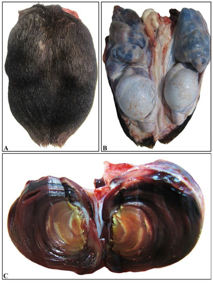
Veterinary Sciences | Free Full-Text | Varicocele in an Adult ...
Sheep Brain Label | Brain anatomy, Dissection, Human brain diagram Sheep Brain Label A drawing of the brain with the parts unlabeled. Students can practice naming the parts of the brain, then check their answers with the provided key. Biologycorner 17k followers More information unlabeled brain Find this Pin and more on A&P by Dijana Kovacevic. Human Brain Diagram Brain Gym For Kids Brain Anatomy And Function
Sheep Brain
Sheep Brain Dissection Project Guide | HST Learning Center Place the brain with the curved top side of the cerebrum facing up. Use a scalpel (or sharp, thin knife) to slice through the brain along the center line, starting at the cerebrum and going down through the cerebellum, spinal cord, medulla, and pons. Separate the two halves of the brain and lay them with the inside facing up. 2.

PPT - Lab Ex. 29 Sheep Brain PowerPoint Presentation, free ...
Sheep Brain - Dorsal View The rostral colliculus(large arrow label) and the caudal colliculus(small arrow label) together form the tectumof the midbrain. Also labeled are the pineal body(green), the caudate nucleus(1), the floor of the fourth ventricle(white and pink) and cerebellar peduncles(blue = rostral, red = middle, and yellow = caudal). Go Top
Brain Dissection - Our Own Secret
Sheep Brain - midsagittal Flashcards | Quizlet Exam 1: Brain Models and Sheep Brain III. 33 terms. kyle_thun. Cerebrum. 22 terms. Jessica_Turner51 PLUS. Respiratory. 47 terms. dezirae-brian_hays. Anatomy Lab: Nervous System. 86 terms. sreisaacs. Other sets by this creator. Intro to Oncology - Drugs. 21 terms. ryankidd01. STLCOP - APPE 2016 - Unit #9 Brand Name. 20 terms.
Warung Makan Mbak Yam, Jebres - GoFood

Sheep brain images | Lab | Amherst College

Close up of a sheep brain cut in half Stock Photo - Alamy
Jual beef slice SUKIYAKI PREMIUM,"PROMO" Indonesia|Shopee ...
Sheep Brain Dissection

Biopsych Brain Dissection | Dissection of a sheep's brain in ...
Biology 7 - BIOLOGY JUNCTION
Sheep Cranial Nerve Roots

Exif | Dorsal Sheep Brain 1 (unlabeled) | Flickr - Photo Sharing!

Close up of a sheep brain cut in half Stock Photo - Alamy

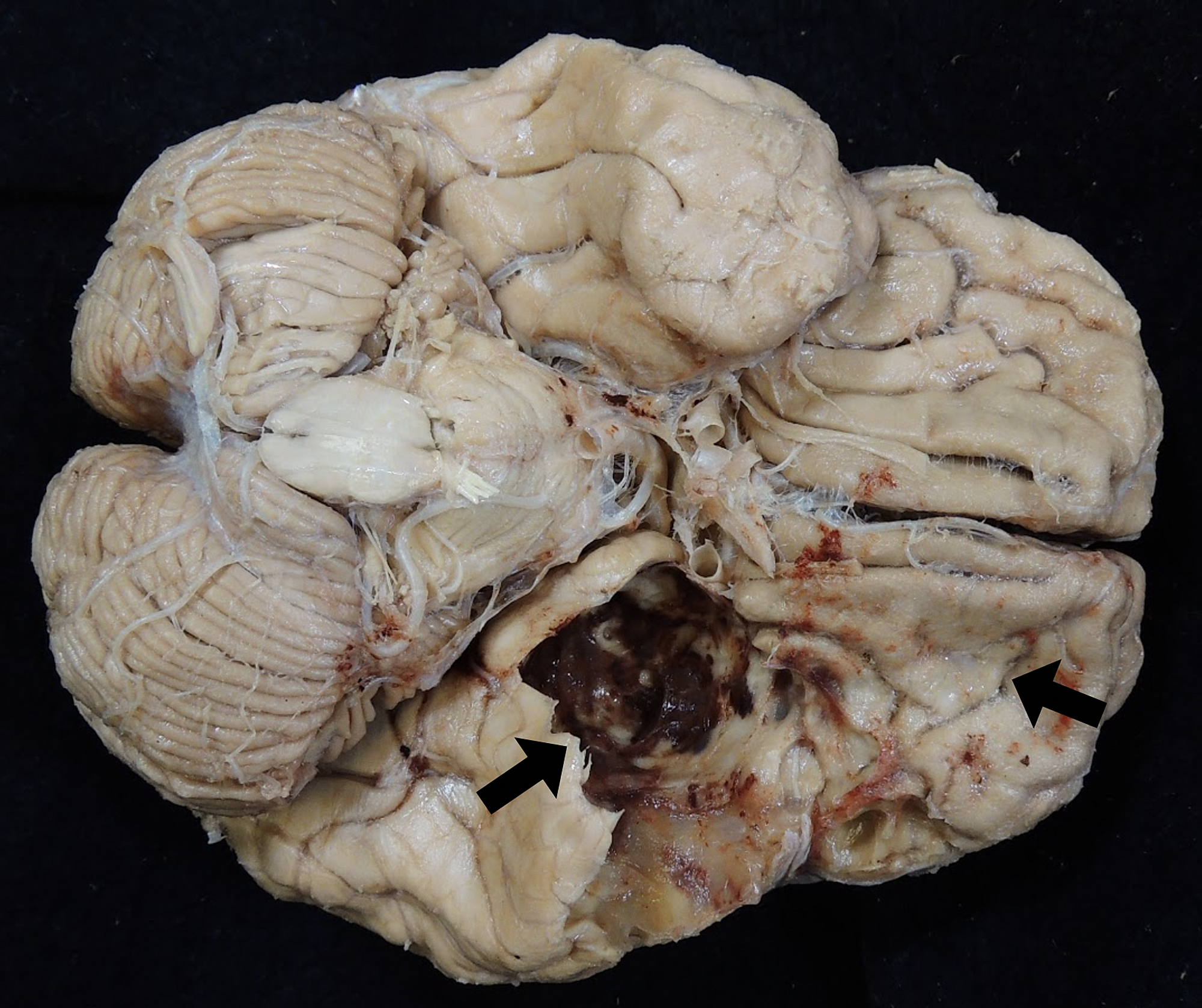
The inferior surface of the left frontal lobe was affected by ...

Sheep Brain Images
STANDARD OPERATING PROCEDURE:

Pig Brain Dissection Worksheet | The Biology of Thought ...
Sheep Brain Neuroanatomy Online Self-Test | KPU.ca - Kwantlen ...
Cureus | Gross and Histological Examination of a Large Spheno ...
Vak Vektor Stok, Ilustrasi Vak Bebas Royalti - Halaman 55 ...
Sheep Brain Dissection

BIO201-Sheep Brain

Vista medial do hemisfério cerebral esquerdo de quati : a ...

Sheep brain & eye (Practical 3) Flashcards | Quizlet
Inferior View of the Optic Radiations | Neuroanatomy | The ...

Sheep Brain Images

Sheep Brain
Post a Comment for "38 sheep brain unlabeled"