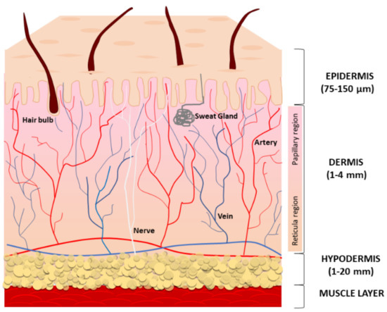
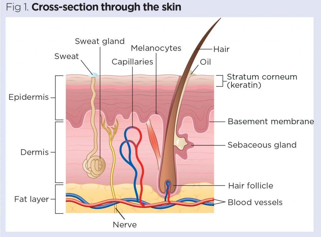
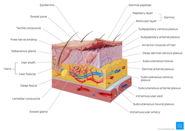
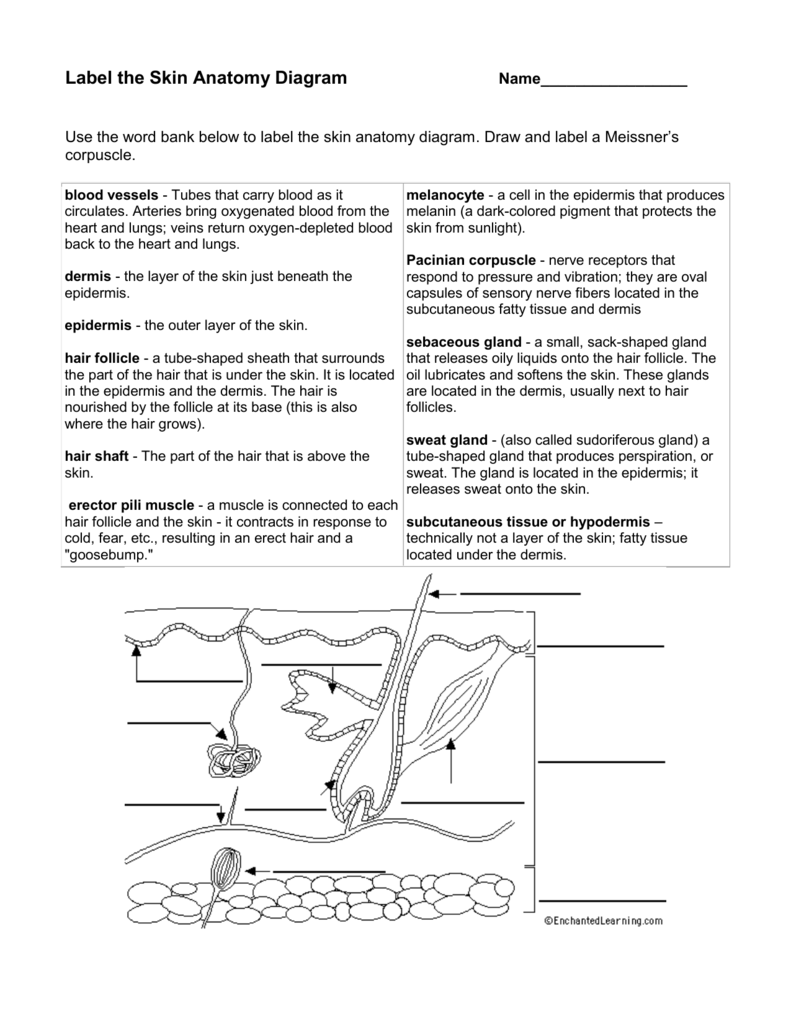
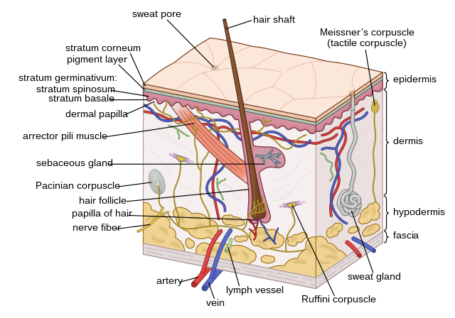
45 label the skin structure and areas indicated in the accompanying diagram of skin
Picture of the Skin - WebMD The skin is the largest organ of the body, with a total area of about 20 square feet. The skin protects us from microbes and the elements, helps regulate body temperature, and permits the... Label the Skin | Biology Diagram | Quizlet Epidermis. layer of the skin that is continually being shed to make room for new skin. Dermis. layer of the skin that contains nerves, vessels, hair follicles, glands, and muscles. Hypodermis. layer of the skin that is used for fat storage. Sweat Pore. opening in the skin that sweat comes out of. Hair Follicle.
Oregon Secretary of State Administrative Rules (a) All public and private enclosed areas at the location that are used in the business operated at the location, including offices, kitchens, rest rooms and storerooms; (b) All areas outside a building that the Commission has specifically licensed for the production, processing, wholesale sale or retail sale of marijuana items; and

Label the skin structure and areas indicated in the accompanying diagram of skin
Solved tive tissue 4. Label the skin structures and areas - Chegg Label the skin structures and areas indicated in the accompanying diagram of thin skin. Then, complete the statements that follow. Weisshaft Stratum opidamist Stratum Stratum Stratum Papilary layer Dermis Reticular layer ascectors allmusde Encrine Sweet blond Dermal Vascular plexus pensery neare fiber Blood vessel Subcutaneous tissue or PDF Integumentary Review Sheet - Home - Holly H. Nash-Rule, PhD Name Lab Time/Date The Integumentary System Basic Structure of the Skin 1. Complete the following statements by writing the appropriate word or phrase on the correspondingly numbered blank: The two basic tissues of which the skin is composed are dense irregular 1. connective tissue, which makes up the dermis, and 1 , which forms the epidermis. Chernobyl disaster - Wikipedia The test was to be conducted during the day-shift of 25 April 1986 as part of a scheduled reactor shut down. The day shift crew had been instructed in advance on the reactor operating conditions to run the test and in addition, a special team of electrical engineers was present to conduct the one-minute test of the new voltage regulating system once the correct conditions had been reached.
Label the skin structure and areas indicated in the accompanying diagram of skin. 2011 ASHRAE HANDBOOK HVAC Applications SI Edition - Academia.edu Enter the email address you signed up with and we'll email you a reset link. 13. CHAPTER XIII. INTEGUMENTARY SYSTEM_LAB.docx - ROJO,... View 13. CHAPTER XIII. INTEGUMENTARY SYSTEM_LAB.docx from BSN 101 at University of Baguio. ROJO, SAMANTHA ISABELLE LABORATORY: Activity: A. Label the skin structures and areas indicated in the PDF 1.Base your answer to the following question on Which 3.The diagram ... Base your answers to questions 34 and 35 on the information and diagram below and on your knowledge of biology. A small water plant (elodea) was placed in bright sunlight for five hours as indicated below. Bubbles of oxygen gas were observed being released from the plant. A)producing sugar B)making protein Label Skin Diagram Printout - EnchantedLearning.com epidermis - the outer layer of the skin. hair follicle - a tube-shaped sheath that surrounds the part of the hair that is under the skin. It is located in the epidermis and the dermis. The hair is nourished by the follicle at its base (this is also where the hair grows). hair shaft - The part of the hair that is above the skin.
Clinical Instructions for Using Silver Diamine Fluoride (SDF ... Condition the lesion and surrounding areas with 20% polyacrylic acid for 10 seconds (removing the smear layer and activating the surface for ionic exchange). It is important to condition not just the lesion but the surrounding areas as well. 5. Rinse with water for 10 seconds and blot dry (leaving a moist "glossy" surface). 6. Solved > 11.The terms and phrases in the key relate:1608365 ... | ScholarOn 11.The terms and phrases in the key relate to the muscle tissues. For each of the three muscle tissues, select the terms or phrases that characterize it, and write the corresponding letter of each term on the answer line. Key: a.striated. b.branching cells. The Integumentary System_page2_answers.png - 4. Label the skin ... Label the skin structures and areas indicated in the accompanying diagram of thin skin. Then, complete The Integumentary System_page2_answers.png - 4. Label the... School South University, Savannah Course Title BIO 1012 Uploaded By Xtina0826 Pages 1 This preview shows page 1 out of 1 page. View full document End of preview. PDF Integumentary System Review Sheet Exercise 7 Answers An outer layer of cells designed to provide protection Keratinized stratified squamous epithelium. hypodermis (subcutaneous layer), this is a layer of *fat located under the dermis of the skin....
4. Label the skin structu - YUMPU Label the skin structures and areas indicated in the accompanying diagram of skin. epidermis dermis Subcutaneous tissue or hypodermis Pacinian corpuscle (deep pressure receptor) 5. What substance is manufactured in the skin (but is not a secretion) to play a role elsewhere in the body? The skin is the site of vitamin D synthesis for the body. Diagram of Human Heart and Blood Circulation in It Four Chambers of the Heart and Blood Circulation. The shape of the human heart is like an upside-down pear, weighing between 7-15 ounces, and is little larger than the size of the fist. It is located between the lungs, in the middle of the chest, behind and slightly to the left of the breast bone. The heart, one of the most significant organs ... Houston Community College Label the skin structures and areas indicated in the accompanying diagram of thin skin. Then, complete the statements that follow. Subcutaneous tissue or (deep pressure receptor) the epidermis. Stratum Stratum Stratum Stratum (layers) Papillary layer Reticular layer Blood vessel Adipose cells Cranial nerves quizzes and labeling exercises | Kenhub Free labeling quiz. Try to understand and memorize what you can from the labeled diagram, then, try to label the cranial nerves yourself with our cranial nerves labeling quiz exercise available to download below. This is a great way to start to get the cogs turning and warm up your memory before you take our other cranial nerve quizzes (but one ...
Solved > 3.Describe five general characteristics of ... - ScholarOn 3.Using the key choices, choose all responses that apply to the following descriptions. Some terms are used more than once. Key:a.stratum basaled.stratum lucidumg.reticular layer ... 4.Label the skin structures and areas indicated in the accompanying diagram of thin skin. Then, complete the statements that follow. a.
Solved C. Label the skin structure and areas indicated in - Chegg C. Label the skin structure and areas indicated in the accompanying diagram of skin D. Appendages of the Skin Using the key choices, respond to the following descriptions. (Some choices may be used more than once). Key: arrector pili muscle Cutaneous receptors Hair Sweat gland-apocrine Sweat gland-eccrine hair follicle nail sebaceous glands 1.
Living Environment - New York Regents June 2015 Exam Structure A is most likely a (1) mitochondrion that synthesizes food for the cell (2) nucleus that is the site of food storage (3) mitochondrion that absorbs energy from the Sun (4) nucleus that is responsible for the storage of information Answer: 7.
Skin Structure (Labeling) Flashcards | Quizlet Created by SRMccoy Terms in this set (19) Hair Shaft Epidermis Dermis Papillary Layer Reticular Layer Hypodermis Sensory Nerve Fiber Lamellar Corpuscle Hair Follicle Receptor Dermal Papillae Subpapillary Plexus Sweat Pore Eccrine Sweat Gland Arrector Pili Muscle Sebaceous (oil) Gland Recommended textbook solutions
PDF Chapter 3 Review Materials Key - wtps.org 3. Figure 3.3 is a simplified diagram of the plasma membrane. Structure A repre- sents channel proteins constructing a pore, structure B represents an ATP- energized solute pump, and structure C is a transport protein that does not depend on energy from ATP. Identify these structures and the membrane phospholipids by color before continuing.
Cambridge International AS & A Level Biology Coursebook ... Figure 1.4: Structure of a generalised animal cell (diameter about 20 μm) as seen with a very high quality light microscope.-R s-C am br ev ie id g w e Figure 1.4 is a drawing showing the structure of a generalised animal cell and Figure 1.5 is a drawing es w-R am br ev id ie ge cell surface membrane nuclear envelope centriole – always found ...
DOC Name: _______Key____ - Mrs. N. Gill The area enclosed by the body wall of the sponge amebocyte ... Label each structure on the diagram. clockwise from the top: pharynx, esophagus, crop, gizzard, intestine, anus. ... The accompanying diagram shows the internal structures of a representative arthropod, the grasshopper. Refer to the diagram to answer the questions that follow.
DOCX Boyertown Area School District Shoulder blade (scapular) Anterior surface of the knee (patellar)34. Spinal column (vertebral) Leg (crural)35. Back of the elbow (cubital) Ankle (tarsal)36. Buttock (gluteal) Toes (digital)37. Hollow behind knee (popliteal) Forehead (frontal)38. Calf (sural) Eye (orbital)39. Side of the leg (fibural or peroneal) Ear (otic)40. Sole (plantar)
Anatomy and Physiology 2e - 2e - Open Textbook Library Anatomy and Physiology 2e is developed to meet the scope and sequence for a two-semester human anatomy and physiology course for life science and allied health majors. The book is organized by body systems. The revision focuses on inclusive and equitable instruction and includes new student support. Illustrations have been extensively revised to be clearer and more inclusive. The web-based ...
PDF Skin Diagram Worksheet Nov 26, 2020 — Label the diagram in the spaces provided. ... What happens in the skin when blood vessels dilate, and how does this regulate temperature?. The two basic tissues of which the skin is composed are dense irregular ... Label the skin structures and areas indicated in the accompanying diagram of thin ....
The Skin (Integumentary System) - YUMPU 4. Label the skin structures and areas indicated in the accompanying diagram of skin. epidermis. dermis. Subcutaneous. tissue or. hypodermis. Pacinian corpuscle (deep pressure receptor) 5. What substance is manufactured in the skin (but is not a secretion) to play a role elsewhere in the body? The skin is the site of vitamin D synthesis for the ...
Answered: PRE-LAB Activity 6: Exploring the… | bartleby PRE-LAB Activity 6: Exploring the Structure and Function of the Ear 1. Use the list of terms provided to label the accompanying illustration of the ear. Check off each term as you label it. O auricle O incus O stapes cochlea O malleus O tympanic membrane O external auditory canal O semicircular canals O vestibule a b. 17 2.
(PDF) Nanda NIC NOC | dwi adiyanto - Academia.edu Enter the email address you signed up with and we'll email you a reset link.
PDF Label The Skin Anatomy Diagram Answers May 11th, 2018 - The Integumentary System Basic Structure of the Skin 1 Label the skin structures and areas indicated in the accompanying diagram of thin skin''5 2 Accessory Structures of the Skin â€" Anatomy and Physiology May 9th, 2018 - 21 1 Anatomy of the Lymphatic Identify the accessory structures of the skin Describe the structure
PDF Integumentary Review Packet Key - Home - Buckeye Valley OBJECTIVE 1 structure(s) of the in OBJECTIVE 2 6. The layer where the skin is thick, such as the palms of the hands and the soles of the feet, is 10 -12 OBJECTIVE 3 OBJECTIVE 4 OBJECTIVE 5 OBJECTIVE 6 develops from a gro OBJECTIVE 7 c. production of gray pigments. d. reduction of melanocyte activity. 4.
PDF The Integumentary System - Holly H. Nash-Rule, PhD Label the skin structures and areas indicated in the accompanying diagram of thin skin. Then, complete the statements that follow. a. Lamellar granules contain glycolipids that prevent water loss from the skin. b. Fibers in the dermis are produced by fibroblasts . c.
PDF Teacher Instructions - Scholastic access to an outdoor area before 10 a.m. and again between 12 p.m. ... 5th-Grade Extension Have students write an accompanying report about what the skin does, how the sun can affect the skin, and ... You will create a 3-D model of the structure of the skin. Use the checklist to label the diagram below and make sure your model has all the ...
Looking at the Structure of Cells in the Microscope A typical animal cell is 10-20 μm in diameter, which is about one-fifth the size of the smallest particle visible to the naked eye. It was not until good light microscopes became available in the early part of the nineteenth century that all plant and animal tissues were discovered to be aggregates of individual cells. This discovery, proposed as the cell doctrine by Schleiden and Schwann ...
Chernobyl disaster - Wikipedia The test was to be conducted during the day-shift of 25 April 1986 as part of a scheduled reactor shut down. The day shift crew had been instructed in advance on the reactor operating conditions to run the test and in addition, a special team of electrical engineers was present to conduct the one-minute test of the new voltage regulating system once the correct conditions had been reached.
PDF Integumentary Review Sheet - Home - Holly H. Nash-Rule, PhD Name Lab Time/Date The Integumentary System Basic Structure of the Skin 1. Complete the following statements by writing the appropriate word or phrase on the correspondingly numbered blank: The two basic tissues of which the skin is composed are dense irregular 1. connective tissue, which makes up the dermis, and 1 , which forms the epidermis.
Solved tive tissue 4. Label the skin structures and areas - Chegg Label the skin structures and areas indicated in the accompanying diagram of thin skin. Then, complete the statements that follow. Weisshaft Stratum opidamist Stratum Stratum Stratum Papilary layer Dermis Reticular layer ascectors allmusde Encrine Sweet blond Dermal Vascular plexus pensery neare fiber Blood vessel Subcutaneous tissue or





(118).jpg)










Post a Comment for "45 label the skin structure and areas indicated in the accompanying diagram of skin"