38 diagram of a microscope with labels
Microscope Parts, Function, & Labeled Diagram - slidingmotion Microscope parts labeled diagram gives us all the information about its parts and their position in the microscope. Microscope Parts Labeled Diagram The principle of the Microscope gives you an exact reason to use it. It works on the 3 principles. Magnification Resolving Power Numerical Aperture. Parts of Microscope Head Base Arm Eyepiece Lens Parts of the Microscope with Labeling (also Free Printouts) Parts of the Microscope with Labeling (also Free Printouts) By Editorial Team March 7, 2022 A microscope is one of the invaluable tools in the laboratory setting. It is used to observe things that cannot be seen by the naked eye. Table of Contents 1. Eyepiece 2. Body tube/Head 3. Turret/Nose piece 4. Objective lenses 5. Knobs (fine and coarse) 6.
Label the microscope — Science Learning Hub In this interactive, you can label the different parts of a microscope. Use this with the Microscope parts activity to help students identify and label the main parts of a microscope and then describe their functions. Drag and drop the text labels onto the microscope diagram.

Diagram of a microscope with labels
Interactive Bacteria Cell Model - CELLS alive Periplasmic Space: This cellular compartment is found only in those bacteria that have both an outer membrane and plasma membrane (e.g. Gram negative bacteria).In the space are enzymes and other proteins that help digest and move nutrients into the cell. Cell Wall: Composed of peptidoglycan (polysaccharides + protein), the cell wall maintains the overall shape of a … Looking at the Structure of Cells in the Microscope A light microscope. (A) Diagram showing the light path in a compound microscope. Light is focused on the specimen by lenses in the condensor. ... and fluorescent labels are usually used for the most precise optical localization. Antibodies are made most simply by injecting a sample of the antigen several times into an animal such as a rabbit or ... Assignment Essays - Best Custom Writing Services Get 24⁄7 customer support help when you place a homework help service order with us. We will guide you on how to place your essay help, proofreading and editing your draft – fixing the grammar, spelling, or formatting of your paper easily and cheaply.
Diagram of a microscope with labels. Microscope Types (with labeled diagrams) and Functions Simple microscope labeled diagram Simple microscope functions It is used in industrial applications like: Watchmakers to assemble watches Cloth industry to count the number of threads or fibers in a cloth Jewelers to examine the finer parts of jewelry Miniature artists to examine and build their work Also used to inspect finer details on products rsscience.com › stereo-microscopeParts of Stereo Microscope (Dissecting microscope) – labeled ... Labeled part diagram of a stereo microscope Major structural parts of a stereo microscope. There are three major structural parts of a stereo microscope. The viewing Head includes the upper part of the microscope, which houses the most critical optical components, including the eyepiece, objective lens, and light source of the microscope. Microscope label Diagram | Quizlet Start studying Microscope label. Learn vocabulary, terms, and more with flashcards, games, and other study tools. Anatomy System - Human Body Anatomy diagram and chart … Plant Cell Lab Makeup – microscope observation of onion and elodea, if students missed the lab that day they can view a site with pictures to complete lab handout Plant Cell Virtual Lab – use a virtual microscope to view plant cells. ... Cell Picture With Labels Image Diagram - Cell Picture With Labels Image Chart - Human anatomy diagrams ...
Labeling the Parts of the Microscope | Microscope World Resources Labeling the Parts of the Microscope This activity has been designed for use in homes and schools. Each microscope layout (both blank and the version with answers) are available as PDF downloads. You can view a more in-depth review of each part of the microscope here. Download the Label the Parts of the Microscope PDF printable version here. en.wikipedia.org › wiki › Wikipedia:Citation_neededWikipedia:Citation needed - Wikipedia If someone tagged your contributions with a "Citation needed" tag or tags, and you disagree, discuss the matter on the article's talk page.The most constructive thing to do in most cases is probably to supply the reference(s) requested, even if you feel the tags are "overdone" or unnecessary. Single-neuron projectome of mouse prefrontal cortex - Nature Mar 31, 2022 · Prefrontal cortex (PFC) is the cognitive center that integrates and regulates global brain activity. However, the whole-brain organization of PFC axon projections remains poorly understood. Using ... › techniques › multi-photonMultiphoton Microscopy | Nikon’s MicroscopyU This is a valuable enhancement to the capability of the conventional microscope since ultraviolet wavelengths below approximately 300 nanometers are very problematic for regular microscope optics. Higher-order non-linear effects, such as four-photon absorption, have been experimentally demonstrated, although it is unlikely that these phenomena ...
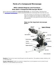
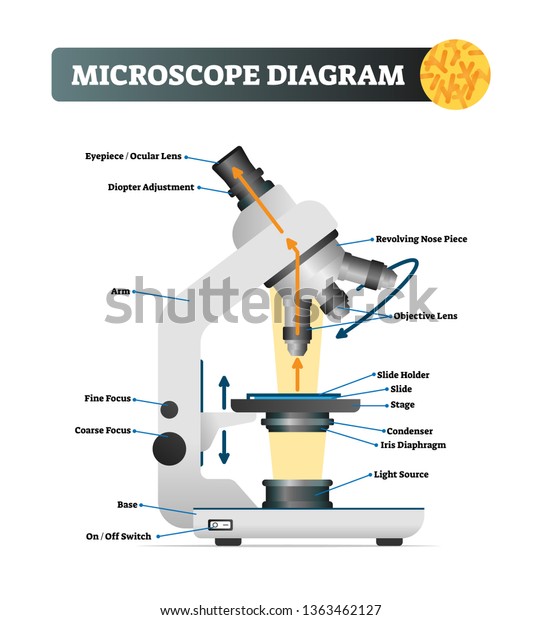
16 Parts of a Compound Microscope: Diagrams and Video Once you have an understanding of the parts of the microscope it will be much easier to navigate around and begin observing your specimen, which is the fun part! The 16 core parts of a compound microscope are: Head (Body) Arm. Base. Eyepiece. Eyepiece tube. Compound Microscope Parts - Labeled Diagram and their Functions Labeled diagram of a compound microscope Major structural parts of a compound microscope There are three major structural parts of a compound microscope. The head includes the upper part of the microscope, which houses the most critical optical components, and the eyepiece tube of the microscope. Microscope Parts and Functions Most specimens are mounted on slides, flat rectangles of thin glass. The specimen is placed on the glass and a cover slip is placed over the specimen. This allows the slide to be easily inserted or removed from the microscope. It also allows the specimen to be labeled, transported, and stored without damage. Two-photon excitation microscopy - Wikipedia Two-photon excitation microscopy (TPEF or 2PEF) is a fluorescence imaging technique that allows imaging of living tissue up to about one millimeter in thickness, with 0.64 μm lateral and 3.35 μm axial spatial resolution. Unlike traditional fluorescence microscopy, in which the excitation wavelength is shorter than the emission wavelength, two-photon excitation requires …
Compound Microscope- Definition, Labeled Diagram, Principle ...
Achiever Papers - We help students improve their academic standing Professional academic writers. Our global writing staff includes experienced ENL & ESL academic writers in a variety of disciplines. This lets us find the most appropriate writer for any type of assignment.

Microscope Diagram - Label Diagram | Quizlet
Compound Microscope Parts, Functions, and Labeled Diagram Compound Microscope Parts, Functions, and Labeled Diagram Posted by Fred Koenig on Nov 18th 2020 Compound Microscope Parts, Functions, and Labeled Diagram Parts of a Compound Microscope Each part of the compound microscope serves its own unique function, with each being important to the function of the scope as a whole.
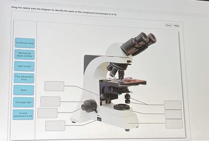
Solved Drag the labels onto the diagram to identify the ...
anatomysystem.comAnatomy System - Human Body Anatomy diagram and chart images ... Cell Picture With Labels Image Diagram - Cell Picture With Labels Image Chart - Human anatomy diagrams and charts explained. This anatomy system diagram depicts Cell Picture With Labels Image with parts and labels. Best diagram to help learn about health, human body and medicine.

Label a Microscope Worksheet
A Study of the Microscope and its Functions With a Labeled Diagram ... To better understand the structure and function of a microscope, we need to take a look at the labeled microscope diagrams of the compound and electron microscope. These diagrams clearly explain the functioning of the microscopes along with their respective parts. Man's curiosity has led to great inventions. The microscope is one of them.

List: Parts of a Microscope and their Function | Pathwooded
Microscope Diagram - Label Diagram | Quizlet The bottom of the microscope, used for support. ocular lens. Eyepiece of a microscope. Diaphragm. Regulates the amount of light on the specimen. nosepiece of microscope. holds the objective lenses. objective lens. The lens on a light microscope that is closest to the stage.
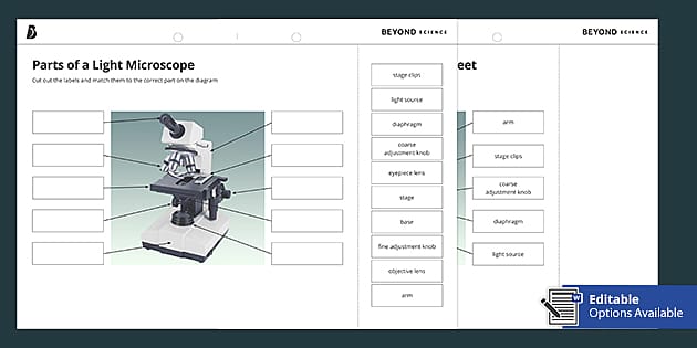
Parts of a Light Microscope Cut and Stick Worksheet - Twinkl
Microscope, Microscope Parts, Labeled Diagram, and Functions The description given below summarize the brief description of microscope parts used to visualize the microscopic specimens such as animal cells, plant cells, microbes, bacteria, viruses, microorganisms etc. The Microscopes parts divided into three different structural parts Head, Base, and Arms.

Lable the microscope worksheet
Parts of a microscope with functions and labeled diagram - Microbe Notes Q. List down the 18 parts of a Microscope. 1. Ocular Lens (Eye Piece) 2. Diopter Adjustment 3. Head 4. Nose Piece 5. Objective Lens 6. Arm (Carrying Handle) 7. Mechanical Stage 8. Stage Clip 9. Aperture 10. Diaphragm 11. Condenser 12. Coarse Adjustment 13. Fine Adjustment 14. Illuminator (Light Source) 15. Stage Controls 16. Base 17.

Simple Microscope Diagram, Formula, Definition, Discoverd by
Compound Microscope Parts, Functions, and Labeled Diagram Nov 18, 2020 · Parts of a Compound Microscope Each part of thenbsp compound microscope serves its own unique function, with each being important to the function of the scope as a whole. The individual parts of a compound microscope can vary heavily depending on the configuration & applications that the scope is being used for. Common compound microscope parts include: …
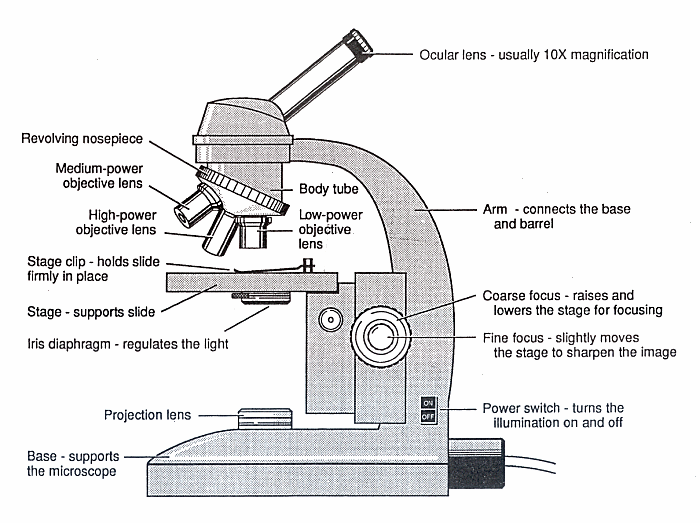
Diagram of a Microscope - Guide to using a microscope
Multiphoton Microscopy | Nikon’s MicroscopyU Two-photon excitation microscopy (also referred to as non-linear, multiphoton, or two-photon laser scanning microscopy) is an alternative to confocal and deconvolution microscopy that provides distinct advantages for three-dimensional imaging.In particular, two-photon excitation excels at imaging of living cells, especially within intact tissues such as brain slices, embryos, whole …

Compound Microscope Parts – Labeled Diagram and their ...
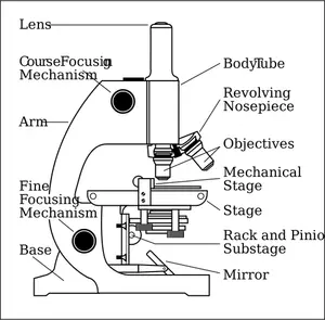
Microscope labeled diagram - SlideShare Follow 1. The Microscope Image courtesy of: Microscopehelp.com Basic rules to using the microscope 1. You should always carry a microscope with two hands, one on the arm and the other under the base. 2. You should always start on the lowest power objective lens and should always leave the microscope on the low power lens when you finish using it.
Glossary of terms used in microscopy – Quekett Microscopical Club
PDF Parts of a Microscope Printables - Homeschool Creations Label the parts of the microscope. You can use the word bank below to fill in the blanks or cut and paste the words at the bottom. Microscope Created by Jolanthe @ HomeschoolCreations.net. Parts of a eyepiece arm stageclips nosepiece focusing knobs illuminator stage objective lenses

A Study of the Microscope and its Functions With a Labeled ...
› createJoin LiveJournal Password requirements: 6 to 30 characters long; ASCII characters only (characters found on a standard US keyboard); must contain at least 4 different symbols;

File:Labelledmicroscope.gif - Wikibooks, open books for an ...
quizlet.com › 231339270 › chapter-1-flash-cardsChapter 1 Flashcards | Quizlet Study with Quizlet and memorize flashcards containing terms like Physiology is to _____ as anatomy is to _____., Which of the following is arranged in correct order from the simplest to the most complex?, The study of tissues with a microscope is called _____. and more.

Microscope - Label - Part 2 Diagram | Quizlet
Compound Microscope - Diagram (Parts labelled), Principle and Uses See: Labeled Diagram showing differences between compound and simple microscope parts Structural Components The three structural components include 1. Head This is the upper part of the microscope that houses the optical parts 2. Arm This part connects the head with the base and provides stability to the microscope.
Microscope drawing - Teaching resources
microscopewiki.com › simple-microscopeSimple Microscope - Diagram (Parts labelled), Principle ... Oct 01, 2022 · The arm of a simple microscope is made of metal and it connects the base of the microscope with the lens tube that houses the eyepiece. Stage . This is a rectangular metal plate with a hole in the middle and is attached to the body of the microscope. The hole in the middle is called the aperture and it allows the light to fall on the specimen.

Microscope Labelling Review Diagram | Quizlet
Microscope: Parts Of A Microscope With Functions And Labeled Diagram. Figure: A diagram of a microscope's components. The microscope has three basic components: the head, the base, and the arm. Head:Occasionally, the head is considered the body. It holds the optical components of the upper part of the microscope. Base:The microscope's base provides great support.
MICROBIO 16 Parts of a Compound Microscope with Diagram and ...
Fluorescence - Wikipedia Fluorescence is the emission of light by a substance that has absorbed light or other electromagnetic radiation.It is a form of luminescence.In most cases, the emitted light has a longer wavelength, and therefore a lower photon energy, than the absorbed radiation.A perceptible example of fluorescence occurs when the absorbed radiation is in the ultraviolet …

Microscope - diagram Tom Butler | Microscope parts, Science ...
Assignment Essays - Best Custom Writing Services Get 24⁄7 customer support help when you place a homework help service order with us. We will guide you on how to place your essay help, proofreading and editing your draft – fixing the grammar, spelling, or formatting of your paper easily and cheaply.

Parts of a Microscope - SmartSchool Systems
Looking at the Structure of Cells in the Microscope A light microscope. (A) Diagram showing the light path in a compound microscope. Light is focused on the specimen by lenses in the condensor. ... and fluorescent labels are usually used for the most precise optical localization. Antibodies are made most simply by injecting a sample of the antigen several times into an animal such as a rabbit or ...

Compound Microscope: Parts of Compound Microscope
Interactive Bacteria Cell Model - CELLS alive Periplasmic Space: This cellular compartment is found only in those bacteria that have both an outer membrane and plasma membrane (e.g. Gram negative bacteria).In the space are enzymes and other proteins that help digest and move nutrients into the cell. Cell Wall: Composed of peptidoglycan (polysaccharides + protein), the cell wall maintains the overall shape of a …

Compound Microscope: Know Definition,working, diagram, properties

Microscope Labeling

Compound Microscope Parts, Diagram Definition, Application ...

Label the microscope — Science Learning Hub

Compound and Stereo- microscopes - Microscopes 4 Schools

Compound Microscope Parts, Functions, and Labeled Diagram ...
Vektor Stok Microscope Diagram Vector Illustration Labeled ...

How to draw compound of Microscope easily - step by step
Mikroskop sisi vektor menggambar dengan bagian-bagian yang ...

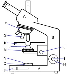
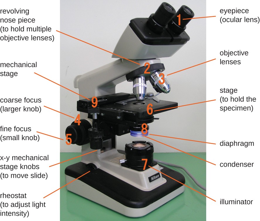
This is a common compound microscope. Label its parts from A ...

Compound Microscope Parts

Microscope Diagram Labeled, Unlabeled and Blank | Parts of a ...

Microscope With Labels clip art | Microscope parts ...

File:Microscope diagram.png - Wikimedia Commons
Instruments of Microscopy | Microbiology | | Course Hero

Label the Microscope Diagram | Download Scientific Diagram

The Microscope
Microscope labeled diagram
Post a Comment for "38 diagram of a microscope with labels"