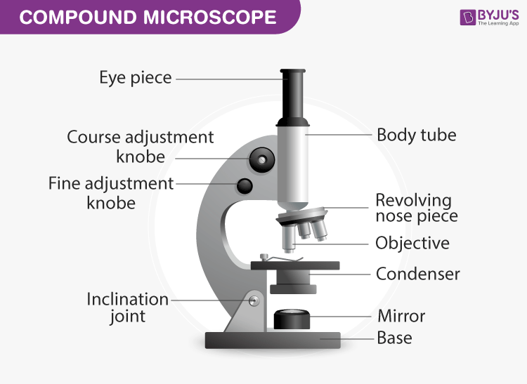
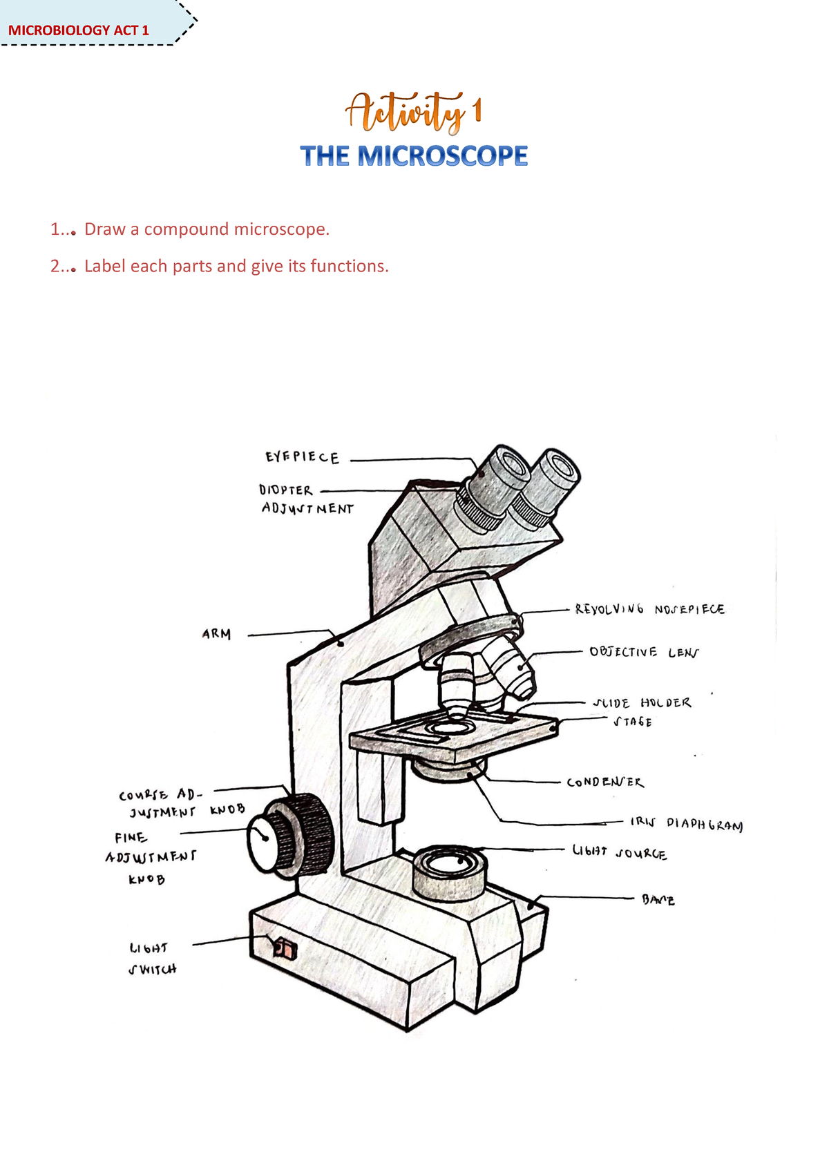
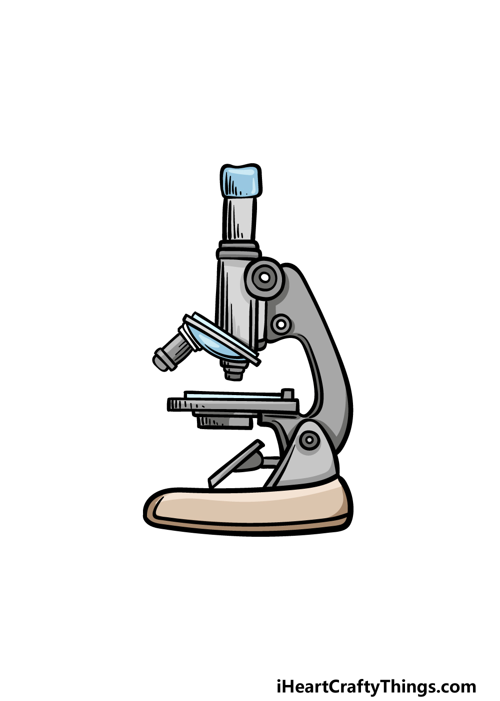
42 drawing of microscope and label
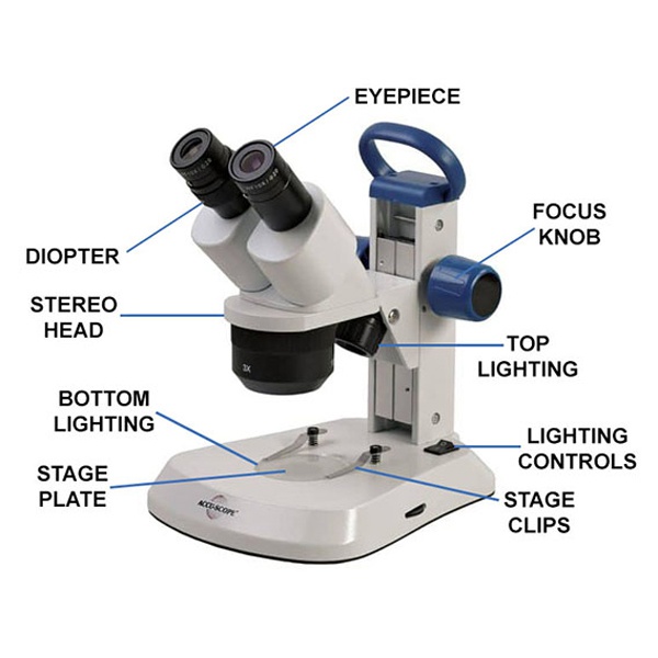
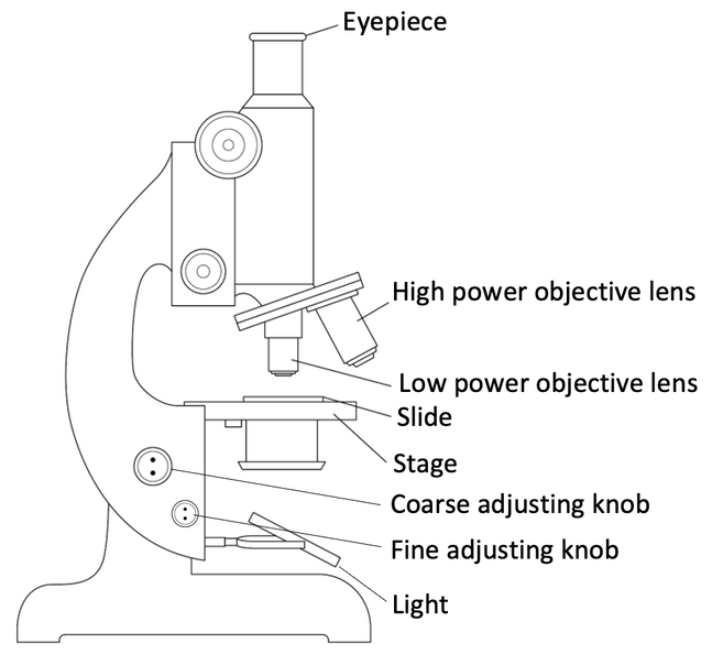
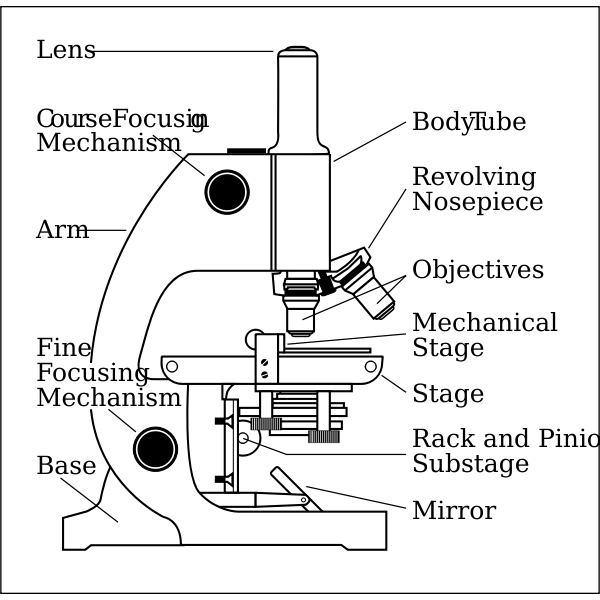
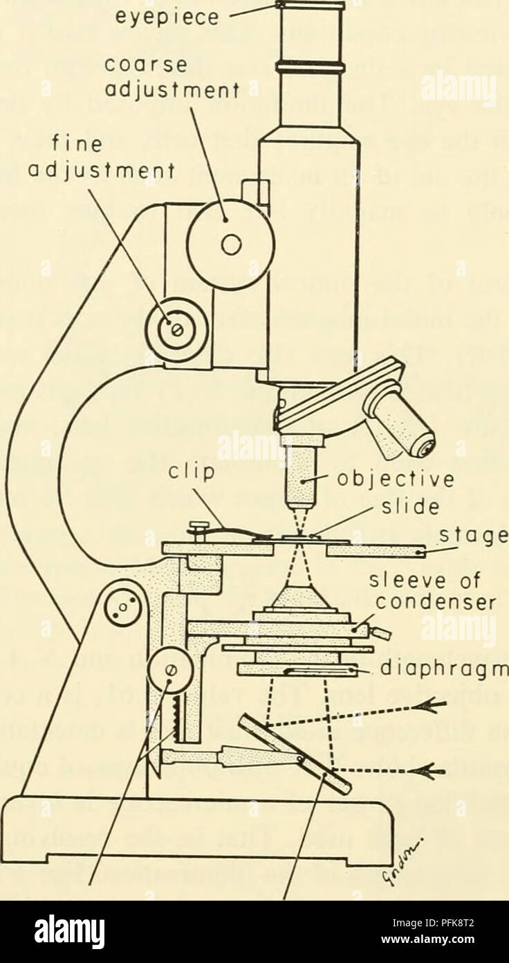
Labelled Diagram of Compound Microscope The below mentioned article provides a labelled diagram of compound microscope. Part # 1. The Stand: The stand is made up of a heavy foot which carries a curved inclinable limb or arm bearing the body tube. The foot is generally horse shoe-shaped structure (Fig. 2) which rests on table top or any other surface on which the microscope in kept. Label Microscope Diagram - EnchantedLearning.com Label Microscope Diagram Using the terms listed below, label the microscope diagram. Inventions and Inventors arm - this attaches the eyepiece and body tube to the base. base - this supports the microscope. body tube - the tube that supports the eyepiece. coarse focus adjustment - a knob that makes large adjustments to the focus.
Lifestyle | Daily Life | News | The Sydney Morning Herald The latest Lifestyle | Daily Life news, tips, opinion and advice from The Sydney Morning Herald covering life and relationships, beauty, fashion, health & wellbeing
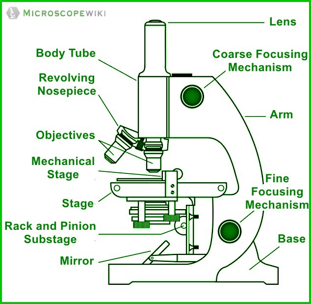
Drawing of microscope and label
Microscope Drawing | How To Draw A Microscope Diagram | Easy ... - YouTube How to draw a microscope diagram. Microscope drawing. Easy and simple step by step tutorial for beginners.Thanks for Watching and Subscribing "Minutes Draw"M... How to Draw a Microscope - Step by Step Easy Drawing Guides Start with the base of the microscope. 2. Sketch the platform of this microscope. 3. Draw the arm of this microscope. 4. Draw the tube of this microscope. 5. Draw the eyepiece for this microscope. Microscope Drawing: How to Sketch Microscope Slides How to Draw Microscope Slides Organize and orient your field of view: To begin, draw a circle as large as possible with a pencil. An 8.5 x 11-inch piece of paper is good size for beginners. The circle represents what you see through the eyepiece of the microscope. Using thin lines, divide the circle into quarters in order to organize the picture.
Drawing of microscope and label. Amazon Coupon Codes: December 2022 Promo Codes 28.11.2022 · Save $15 at Amazon with coupon code citi20... (click to reveal full code). 918 other Amazon coupons and deals also available for November 2022. No Longer Available - KETV Hearst Television participates in various affiliate marketing programs, which means we may get paid commissions on editorially chosen products purchased through our links to retailer sites. How to Sketch a Microscope Slide - Identifying and Sketching Cell ... First, to represent the microscope field of view, draw a circle on the page - this can be freehand or, if you want to be precise, use a compass. If you are using a graticule slide (a microscope slide with millimeter grid lines), lightly sketch a grid over your circle. Microscope, Microscope Parts, Labeled Diagram, and Functions Revolving Nosepiece or Turret: Turret is the part of the microscope that holds two or multiple objective lenses and helps to rotate objective lenses and also helps to easily change power. Objective Lenses: Three are 3 or 4 objective lenses on a microscope. The objective lenses almost always consist of 4x, 10x, 40x and 100x powers. The most common eyepiece lens is 10x and when it coupled with ...
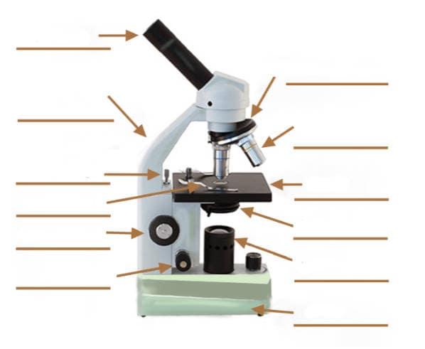
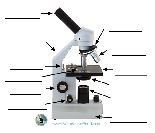
Cyber Monday 2022 | Amazon.com Cyber Monday is an Amazon deals event on November 26-28. Go to Amazon and prepare your holidays with epic deals from top brands on this seasons’ must-have items. Microscope Labeling - The Biology Corner Students label the parts of the microscope in this photo of a basic laboratory light microscope. Can be used for practice or as a quiz. ... Microscope Labeling . Microscope Use: 15. When focusing a specimen, you should always start with the _____ objective. 16. When using the high power objective, only the _____ knob should be used. 17. The ... microscope worksheet label Microscope drawing science diagram biology simple microscopes labeled labeling worksheet parts tom butler technical tools classroom clipart lessons teaching found. Microscope worksheet pdf. ... Parts Of A Microscope Labeling & Functions Worksheet - Science . microscope worksheet parts labeling science. Pin En Ciencia Y Naturaleza No Longer Available - WMUR Hearst Television participates in various affiliate marketing programs, which means we may get paid commissions on editorially chosen products purchased through our links to retailer sites.
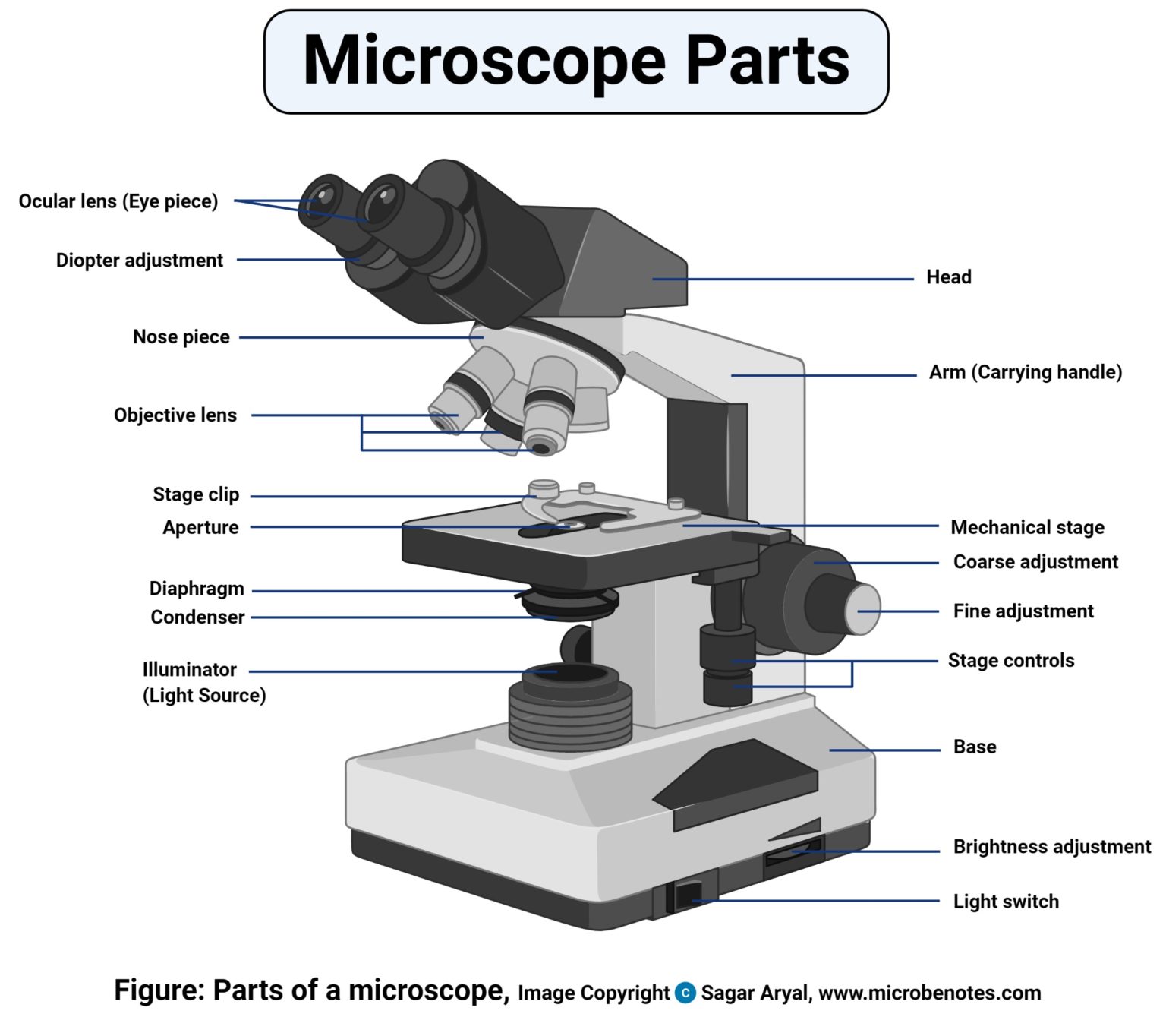
Parts of a microscope with functions and labeled diagram - Microbe Notes Figure: Diagram of parts of a microscope There are three structural parts of the microscope i.e. head, base, and arm. Head - This is also known as the body. It carries the optical parts in the upper part of the microscope. Base - It acts as microscopes support. It also carries microscopic illuminators. Microscope Parts LabelingPaul - Biology: Biology Notes/Charts 2015-2016 ... 1 Draw and label the parts of the microscope2. microscope compound drawing parts label light sketch easy diagram drawn lable paintingvalley explain draw drawings pencil line structure its. Students label the parts of the microscope in this photo of a basic laboratory light microscope. You should carry the microscope by the _____ and the _____. › shows › fox-filesFox Files | Fox News Jan 31, 2022 · FOX FILES combines in-depth news reporting from a variety of Fox News on-air talent. The program will feature the breadth, power and journalism of rotating Fox News anchors, reporters and producers. learn.genetics.utah.edu › content › cellsCell Size and Scale - University of Utah Smaller cells are easily visible under a light microscope. It's even possible to make out structures within the cell, such as the nucleus, mitochondria and chloroplasts. Light microscopes use a system of lenses to magnify an image. The power of a light microscope is limited by the wavelength of visible light, which is about 500 nm.
Drawing Of A Microscope And Label - Warehouse of Ideas Here presented 54+ microscope drawing and label images for free to download, print or share. Title Is Informative, Centered, And Larger Than Other Text. How to draw a microscope and label. Compound microscopes have furthered medical research, helped to solve crimes, and they have repeatedly proven invaluable in unlocking the secrets of the.
Cell Size and Scale - University of Utah Smaller cells are easily visible under a light microscope. It's even possible to make out structures within the cell, such as the nucleus, mitochondria and chloroplasts. Light microscopes use a system of lenses to magnify an image. The power of a light microscope is limited by the wavelength of visible light, which is about 500 nm. The most powerful light microscopes can …
A Study of the Microscope and its Functions With a Labeled Diagram ... A Study of the Microscope and its Functions With a Labeled Diagram To better understand the structure and function of a microscope, we need to take a look at the labeled microscope diagrams of the compound and electron microscope. These diagrams clearly explain the functioning of the microscopes along with their respective parts.
Microscope Parts and Functions Most specimens are mounted on slides, flat rectangles of thin glass. The specimen is placed on the glass and a cover slip is placed over the specimen. This allows the slide to be easily inserted or removed from the microscope. It also allows the specimen to be labeled, transported, and stored without damage.
Givenchy official site Discover all the collections by Givenchy for women, men & kids and browse the maison's history and heritage
Compound Microscope Parts - Labeled Diagram and their Functions Labeled diagram of a compound microscope Major structural parts of a compound microscope There are three major structural parts of a compound microscope. The head includes the upper part of the microscope, which houses the most critical optical components, and the eyepiece tube of the microscope.
Microscope Parts, Function, & Labeled Diagram - slidingmotion Microscope parts labeled diagram gives us all the information about its parts and their position in the microscope. Microscope Parts Labeled Diagram The principle of the Microscope gives you an exact reason to use it. It works on the 3 principles. Magnification Resolving Power Numerical Aperture. Parts of Microscope Head Base Arm Eyepiece Lens
Fox Files | Fox News 31.01.2022 · FOX FILES combines in-depth news reporting from a variety of Fox News on-air talent. The program will feature the breadth, power and journalism of rotating Fox News anchors, reporters and producers.
Drawing Of Microscope And Label - Warehouse of Ideas Here presented 54+ microscope drawing and label images for free to download, print or share. All The Best Microscope Drawing 38+ Collected On This Page. Its standard magnification is 10x with an optional eyepiece having.
› CH-FRRéservez des vols pas chers et trouvez des offres ... - easyJet Réservez des vols pas chers sur easyJet.com vers les plus grandes villes d'Europe. Trouvez aussi des offres spéciales sur votre hôtel, votre location de voiture et votre assurance voyage.
Patent Public Search | USPTO Welcome to Patent Public Search. The Patent Public Search tool is a new web-based patent search application that will replace internal legacy search tools PubEast and PubWest and external legacy search tools PatFT and AppFT.
Parts of the Microscope with Labeling (also Free Printouts) Parts of the Microscope with Labeling (also Free Printouts) By Editorial Team March 7, 2022 A microscope is one of the invaluable tools in the laboratory setting. It is used to observe things that cannot be seen by the naked eye. Table of Contents 1. Eyepiece 2. Body tube/Head 3. Turret/Nose piece 4. Objective lenses 5. Knobs (fine and coarse) 6.
Home [ ] Constitutional Rights Foundation (CRF) is a non-profit, non-partisan, community-based organization. CRF seeks to instill in our nation's youth a deeper understanding of citizenship through values expressed in our Constitution and its Bill of Rights and to educate young people to become active and responsible participants in our society.
Microscope Drawing Easy with Label - YouTube Microscope Drawing Easy with Label 886 views Apr 13, 2020 In this video I go over a microscope drawing that is easy with label. There is a blank copy at the end of the video to review...
Labeling the Parts of the Microscope | Microscope World Resources Labeling the Parts of the Microscope This activity has been designed for use in homes and schools. Each microscope layout (both blank and the version with answers) are available as PDF downloads. You can view a more in-depth review of each part of the microscope here. Download the Label the Parts of the Microscope PDF printable version here.
Microscope Drawing: How to Sketch Microscope Slides How to Draw Microscope Slides Organize and orient your field of view: To begin, draw a circle as large as possible with a pencil. An 8.5 x 11-inch piece of paper is good size for beginners. The circle represents what you see through the eyepiece of the microscope. Using thin lines, divide the circle into quarters in order to organize the picture.
How to Draw a Microscope - Step by Step Easy Drawing Guides Start with the base of the microscope. 2. Sketch the platform of this microscope. 3. Draw the arm of this microscope. 4. Draw the tube of this microscope. 5. Draw the eyepiece for this microscope.
Microscope Drawing | How To Draw A Microscope Diagram | Easy ... - YouTube How to draw a microscope diagram. Microscope drawing. Easy and simple step by step tutorial for beginners.Thanks for Watching and Subscribing "Minutes Draw"M...













Post a Comment for "42 drawing of microscope and label"