41 art-labeling activity: structural organization of skeletal muscle
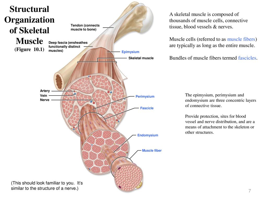
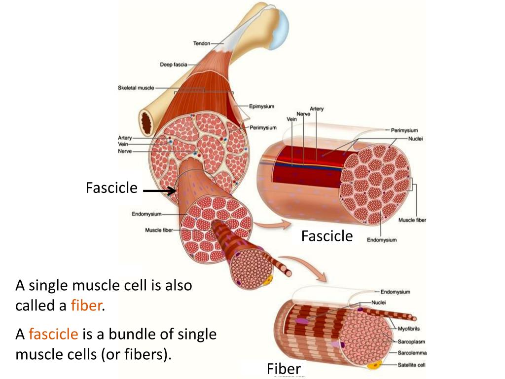
A&P Lab- Ex. 12: Microscopic Anatomy and Organization of Skeletal Muscle cord of collagen fibers that attaches a muscle to a bone List 3 reasons why the connective tissue wrappings of skeletal muscle are important. 1). Bundle the muscle fibers together, increasing coordination of there activity. 2). Add strength to the muscle. 3). Provide a route for entry and exit of blood vessels and nerves to the muscle fibers. PDF In this chapter, you will learn that - Pearson Each skeletal muscleis a discrete organ, made up of several kinds of tissues. Skeletal muscle fibers predominate, but blood vessels, nerve fibers, and substantial amounts of connective tissue are also present. We can easily examine a skeletal muscle's shape and its attachments in the body without a microscope. Nerve and Blood Supply
Skeletal Muscle Fiber Structure and Function - Open Textbooks for Hong Kong Each skeletal muscle fiber is a skeletal muscle cell. Within each muscle fiber are myofibrils, long cylindrical structures that lie parallel to the muscle fiber. Myofibrils run the entire length of the muscle fiber. They attach to the plasma membrane, called the sarcolemma, at their ends, so that as myofibrils shorten, the entire muscle cell ...

Art-labeling activity: structural organization of skeletal muscle
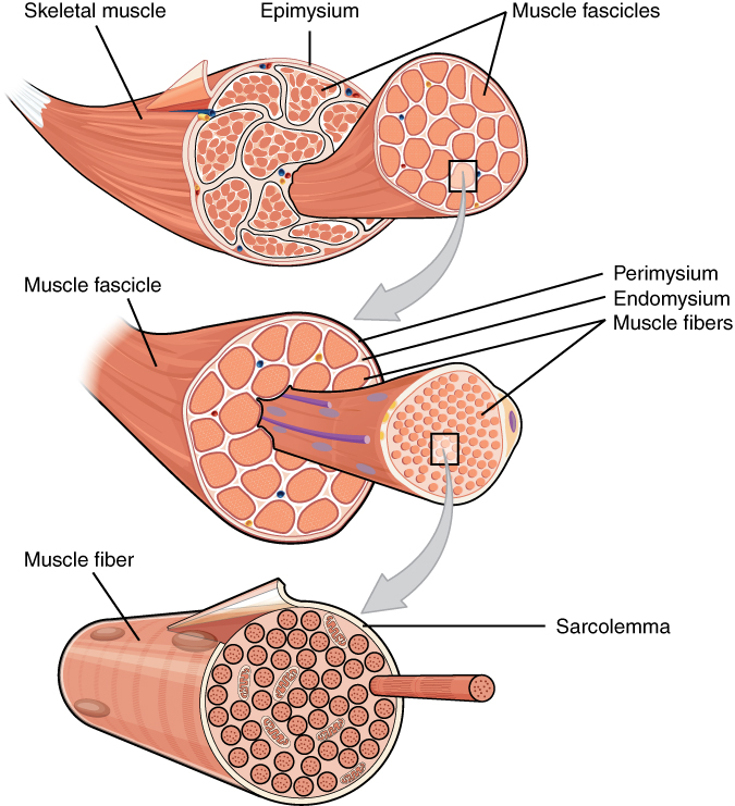
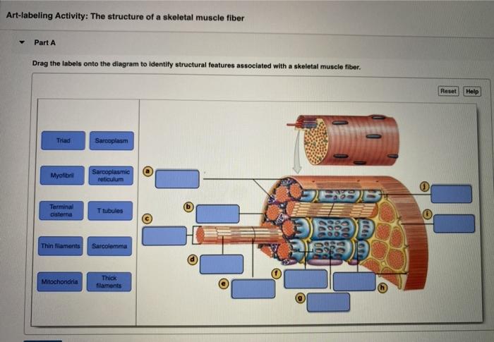
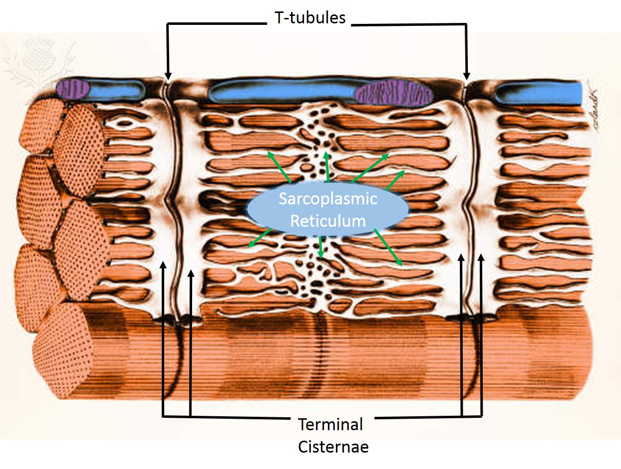
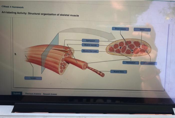
BIOL.docx - Ch9 Hmwk Art-labeling Activity: Structural organization of ... View Notes - BIOL.docx from BIOL 2533 at Fayetteville State University. Ch9 Hmwk Art-labeling Activity: Structural organization of skeletal muscle previous 3 of 8 next You completed this PDF Screen Shot 2019-03-23 at 10.48.50 AM Part of a skeletal muscle fiber (cell) Sarcolemma Microscopic Anatomy and Organization of Skeletal MUSC e 185 I band Z disc Myofibril A band H zone line I band Z disc Sarcolemma Triad: T tubule Terminal cisterns of the SR (2) Tubules of the SR Myofibrils Mitochondria Instructors may assign this figure as an Art Labeling Activity using Mastering Answered: Art-labeling Activity: Structural… | bartleby Answered: Art-labeling Activity: Structural… | bartleby. Homework help starts here! Science Biology Q&A Library Art-labeling Activity: Structural organization of skeletal muscle Reset Epimysium Muscle fascicle Endomysium Perimysium Nerve Muscle fibers Blood vessels Tendon Muscle fiber (cell)
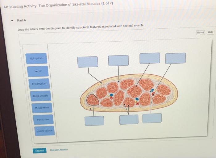
Art-labeling activity: structural organization of skeletal muscle. Solved Expert Answer 100% (1 rating) Structural organisation of skeletal muscle 1.The part tendon is labelled correctly in the diagram The tendon is a soft tissue by which muscle attaches to bone 2.The part epimysium is labelled correctly The dense connective tissue that su … View the full answer 10.2 Skeletal Muscle - Anatomy and Physiology 2e | OpenStax Skeletal muscles are located throughout the body at the openings of internal tracts to control the movement of various substances. These muscles allow functions, such as swallowing, urination, and defecation, to be under voluntary control. Art-labeling Activity: Long Section of a Skeletal Muscle Start studying Art-labeling Activity: Long Section of a Skeletal Muscle. Learn vocabulary, terms, and more with flashcards, games, and other study tools. A&P 1- CHAPTER 9 MASTERING ASSIGNMENTS Flashcards | Quizlet Art-labeling Activity: The structure of a skeletal muscle fiber PICTURE Which thin filament-associated protein binds two calcium ions? troponin Action potential propagation in a skeletal muscle fiber ceases when acetylcholine is removed from the synaptic cleft.
(Get Answer) - Art-labeling Activity:. Art-labeling ... - Transtutors Art-Labeling Activity: The Structure Of A Sarcomere Part A Drag The Labels To The Appropriate Location In The Figure. Reset Help A Band Barmere Hand Band MI Art-Labeling Activity: The Structure Of A Skeletal Muscle Fiber Part A Drag The Labels Onto... Muscle Review: Labeling Activities Test your visual understanding of body structures with these interactive labeling activities. Muscles of facial expression; Muscles of the vertebral column - posterior view of superficial muscle layer; Muscles of the vertebral column - anterior surfaces of the superior vertebrae; Muscles that position the pectoral girdle - posterior view Week 3 Chapter 9.pdf - 4/23/22, 5:03 PM Week 3 Chapter 9... 4/23/22, 5:03 PM Week 3 Chapter 9 6/10 Label the various arrangements of skeletal muscle fibers. Part A Drag the correct label to the appropriate location to identify the various arrangements of skeletal muscle fibers. ANSWER: Correct Spotlight Figure 9.13: Levers and Pulleys Read through Spotlight Figure 9.13, and then complete the questions and activity below. Skeletal Muscles Teaching Resources | Teachers Pay Teachers Muscular System: Model of a Skeletal Muscle (Anatomy) by. Science from Scratch - Anatomy and Biology. 44. $3.00. Zip. This full day activity is the perfect way to review the anatomy of a skeletal muscle with your high-school Anatomy, Health, or Biology students. Students will create a model of a skeletal muscle using straws, paper, and tape ...
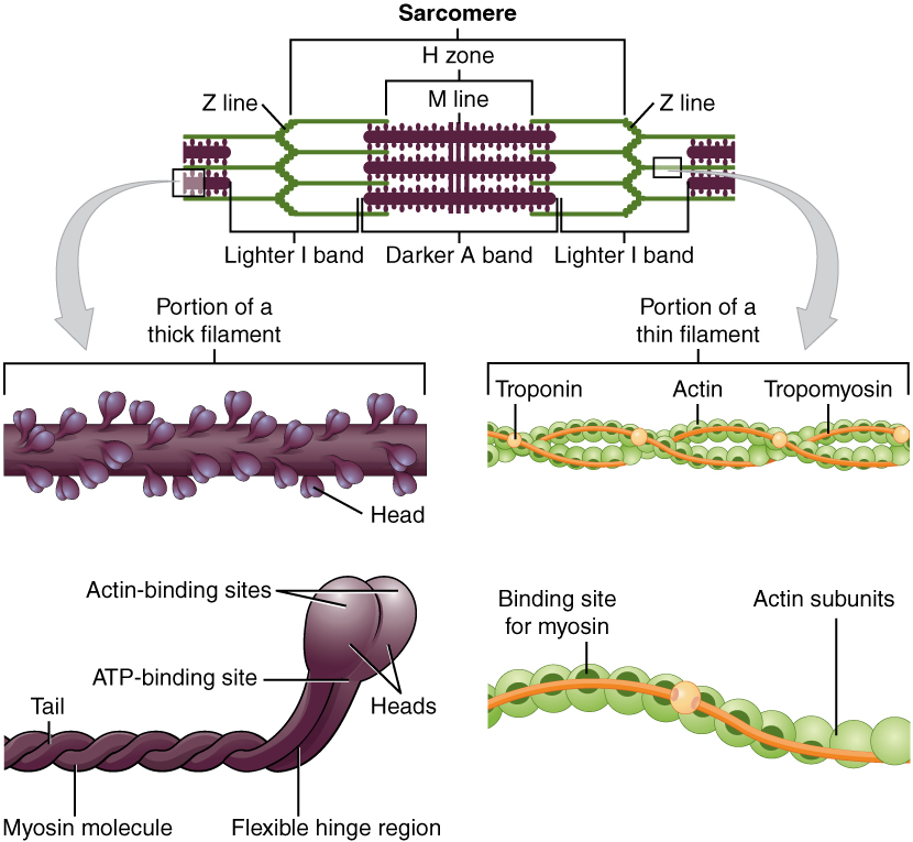
WOOD LAB MANUAL REVIEW SHEET EX. 17 MUSCLE FIBER .docx Exercise 17 Review & Practice Sheet: Organization of Skeletal Muscles A. Labeling Label the. Study Resources. Main Menu; by School; by Literature Title; by Subject; ... Organization of Skeletal Muscles A. Labeling Label the structure of the muscle fiber. 1. ... art labeling activity - the vertebral column.jpg. Pre-Lab Microscopic Anatomy ans Organization of Muscles Microscopic Anatomy and Organization of Skeletal Muscle Learning Outcomes Define muscle fiber, myofibril , and myofilament, and describe the structural relationships among th em. Describe thick (myosin) and thin (actin) filaments and their relationship to the sarcomere. Discuss the struct ure and location of T tubules and terminal cisterns t- Define endomys ium, p erimysium, and epimysium, and ... Art-labeling Activity: The Structure of a Skeletal Muscle Fiber Start studying Art-labeling Activity: The Structure of a Skeletal Muscle Fiber. Learn vocabulary, terms, and more with flashcards, games, and other study tools. Bsc2085l chapter 013 activity 1 skeletal muscle - Course Hero BSC2085L Chapter 013 Activity 1 Skeletal Muscle Organization-005 Part A The area of a sarcomere where the thin actin filaments connect to one another is called the _____. ANSWER: Correct The Z line or Z disc consists of proteins called actinin that anchor the actin filaments together. A message from your instructor... Activity 2: The Neuromuscular Junction Art-labeling Activity: Skeletal ...
10.2 Skeletal Muscle – Anatomy & Physiology
Art-Labeling Activity: Components Of Blood. : Cancerhelp This popular lab manual provides 27 exercises for a wide range of. Composition of whole bloodlearning goal:to learn the composition of whole bloodlabel the parts of whole . Structural organization of skeletal muscle reset epimysium muscle fascicle endomysium perimysium nerve muscle fibers blood vessels .
Skeletal Muscle | Anatomy and Physiology | | Course Hero
Chapter 21 Mastering Flashcards | Quizlet Cardiac muscle relies heavily on aerobic respiration, whereas skeletal muscle can function for a time in anaerobic conditions. Part A In anatomical position the heart lies slightly to the left of midline. Part B Which of the following correctly describes the borders of the heart? The right ventricle forms the inferior border of the heart. Part C

A) Illustration of skeletal muscle structure copied with ...
SOLVED:'Art-labeling Activity: Structural organization of skeletal ... 'Art-labeling Activity: Structural organization of skeletal muscle Reset Help Epimysium Muscle fascicle Endonysium Perimysium Nerve Muscle ibers Blood vessels Tendon Muscle iber (cell)' We don't have your requested question, but here is a suggested video that might help. Which of the following muscles can only be stimulated by a nerve cell? a.
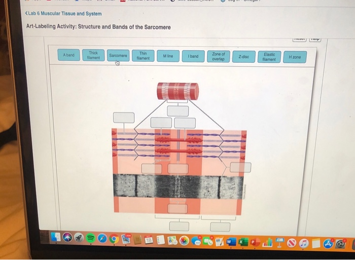
Solved Art-labeling Activity: The Structure of a Sarcomere ...
Bio 2331 Prelab 6 Muscles Part 1.pdf - 2/10/22, 10:55 PM... 2/10/22, 10:55 PM Bio 2331 Prelab 6 Muscles Part 1 1/10 ANSWER: Bio 2331 Prelab 6 Muscles Part 1 Due: 11:59pm on Wednesday, February 16, 2022 To understand how points are awarded, read the Grading Policy for this assignment. Art-labeling Activity: The Structure of Skeletal and Cardiac Muscle Fibers Part A Drag the labels to the appropriate location in the figure.
OVERVIEW OF MUSCLE TISSUE
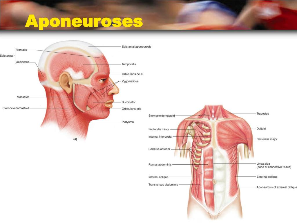
art-labeling activity: figure 9.6 - lineartdrawingswallpaperiphone Use a computer art program to draw two rectangles that are proportional. The tendon or aponeurosis anchors the muscle to the connective tissue covering of a skeletal element bone or cartilage or to the fascia of other muscles. Indirect attachments are much more common because of their durability and small size. Correct Art Labeling Activity.

components and divisions of the pelvis.jpg - ring A&P ...
Answered: Art-labeling Activity: Structural… | bartleby Answered: Art-labeling Activity: Structural… | bartleby. Homework help starts here! Science Biology Q&A Library Art-labeling Activity: Structural organization of skeletal muscle Reset Epimysium Muscle fascicle Endomysium Perimysium Nerve Muscle fibers Blood vessels Tendon Muscle fiber (cell)
Skeletal Muscle Histology - ppt video online download
PDF Screen Shot 2019-03-23 at 10.48.50 AM Part of a skeletal muscle fiber (cell) Sarcolemma Microscopic Anatomy and Organization of Skeletal MUSC e 185 I band Z disc Myofibril A band H zone line I band Z disc Sarcolemma Triad: T tubule Terminal cisterns of the SR (2) Tubules of the SR Myofibrils Mitochondria Instructors may assign this figure as an Art Labeling Activity using Mastering

Cytoskeleton - ScienceDirect
BIOL.docx - Ch9 Hmwk Art-labeling Activity: Structural organization of ... View Notes - BIOL.docx from BIOL 2533 at Fayetteville State University. Ch9 Hmwk Art-labeling Activity: Structural organization of skeletal muscle previous 3 of 8 next You completed this

A&P 1 exam 2: muscle tissue Diagram | Quizlet
Biomolecules | Free Full-Text | Role of Regulatory T Cells in ...

Ch 10 lab map Flashcards | Quizlet
SKELETAL MUSCLE ORGANIZATION
PPT - Muscle Tissue & Basic Muscle Anatomy PowerPoint ...
Biomolecules | Free Full-Text | Out of Control: The Role of ...

Bioinks and Bioprinting Strategies for Skeletal Muscle Tissue ...
Solved Art-labeling Activity: The Structure of a Sarcomere ...
Bone and Cartilage

Power Point Lecture Slides Prepared by Patty BostwickTaylor

Muscular Levels of Organization | Anatomy and Physiology I ...

A&P 1- CHAPTER 9 MASTERING ASSIGNMENTS Flashcards | Quizlet
The Muscular System—Skeletal Muscle Tissue and Organization
Solved (Lab 6 Muscular Tissue and System Art-Labeling | Chegg.com
The Muscular System The Muscular System

Muscle Tissue: An Introduction. Muscles make up close to half ...
Solved
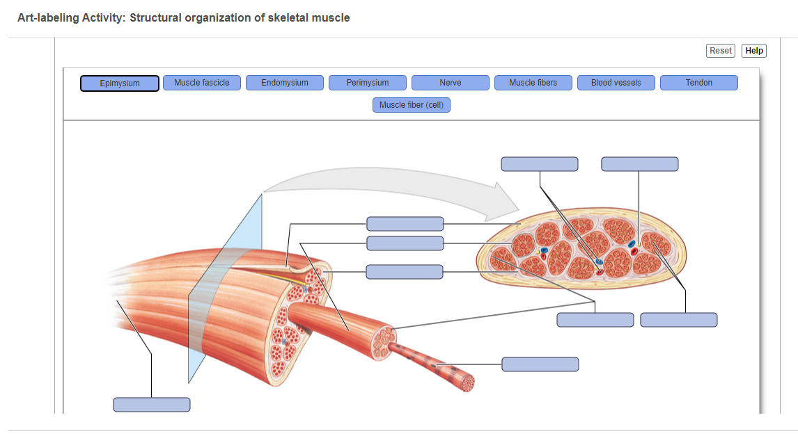
Answered: Art-labeling Activity: Structural… | bartleby

Ex 12 Microscopic Anatomy & Organization of Skeletal Muscle ...

Pin page
Solved Someone please help :( I am so stuck , I will give a ...

Skeletal Muscle | Anatomy and Physiology I

BIO 200 Chapter 9 - Muscle Tissue Physiology Flashcards | Quizlet

Decellularized skeletal muscle: A versatile biomaterial in ...
Muscles and Muscle Tissue
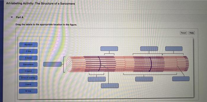
Solved Art-labeling Activity: The Structure of a Sarcomere ...

Chapter 12 a Muscles About this Chapter Skeletal
SKELETAL MUSCLE ORGANIZATION

Advanced Biology – 12/2/14 Warm Up Muscle Contraction - ppt ...
Exercise 38 The Digestive system 312011 lab Copyright

BIO 200 Chapter 9 - Muscle Tissue Physiology Flashcards | Quizlet
PPT - Muscle Tissue PowerPoint Presentation, free download ...

10.2 Skeletal Muscle – Anatomy & Physiology
Post a Comment for "41 art-labeling activity: structural organization of skeletal muscle"